
 Türkçe
 Français
 Deutsch
 Italiano
 Español
 العربية
 Русский
MitraClip is a non-surgical mitral valve regurgitation repair method applied in patients who are at high risk for open-heart surgery, without opening the chest cage and without stopping the heart. Also known as the “clipping” technique in the field of interventional cardiology, this procedure is based on reaching the heart through the femoral vein and fastening the leaking mitral valve leaflets to each other with a special clip. By mechanically correcting severe valve leakage that impairs the heart’s pumping function and reduces quality of life, this technology offers rapid recovery without a surgical incision and significantly reduces heart failure symptoms and life-threatening risks.

Prof. Dr. Kadriye Kılıçkesmez
Cardiology, Interventional Cardiologist – Interventional Cardiologist
Prof. Dr. Kadriye Orta Kılıçkesmez is one of the leading names in the field of Turkish cardiology. She was born on January 24, 1974, in Tekirdağ. After completing her undergraduate education at Istanbul University Cerrahpaşa Faculty of Medicine, she chose cardiology as her specialty and received her specialist training at the Cardiology Institute of the same university.
After working for a short period at Çorlu State Hospital and Turkish Kidney Foundation Service Hospital, she returned to the I.U. Cardiology Institute. Continuing her academic career there, Kadriye Kılıçkesmez became an associate professor in 2012. She then worked at Royal Brompton on complex coronary interventions, CTO intracoronary imaging, and structural heart diseases, and wrote scientific articles. In 2015, she was appointed by the university to establish the Şişli Etfal cardiology clinic and Angio laboratory. Becoming a professor in 2017, Kadriye Kılıçkesmez established the cardiology clinic and Angio laboratory of Prof. Dr. Cemil Taşçıoğlu Hospital in 2020 and ensured that the clinic became a training clinic.
View MoreWhat Is MitraClip (Non-Surgical Mitral Valve Regurgitation Repair)?
MitraClip is a minimally invasive procedure that allows mitral valve regurgitation to be treated without requiring open-heart surgery, using a catheter-based method. Through a catheter advanced from the femoral vein, the mitral valve leaflets are joined with a special clip to reduce backflow. It is particularly preferred in patients with high surgical risk. The procedure is performed under echocardiography guidance, and the recovery time is generally shorter than surgery.
What Does Mitral Valve Regurgitation Change in Our Body?
Understanding how the heart works is the first step to grasping why this treatment is necessary. Our heart is a powerful, four-chambered motor that works nonstop throughout our lives, pumping clean blood to every cell of the body. On the left side of this motor are the left atrium, where clean blood collects, and the left ventricle, which ejects this blood to the body. The mitral valve is a gate located between these two chambers, enabling one-way blood flow.
Each time the heart contracts, this gate must close tightly and prevent blood from leaking backward. However, in mitral regurgitation, this gate cannot close. As a result, a significant portion of the blood that should go to the body flows back toward the lungs, where it came from. This greatly increases the heart’s workload. To meet the body’s needs, the heart has to work harder and beat faster. Over time, the tired heart muscle enlarges, weakens, and loses its pumping function.
This mechanical impairment is reflected very clearly in the patient’s daily life through distinct symptoms. Our patients often say they can no longer do tasks they previously did with ease. Due to the pressure of blood and fluid accumulating in the lungs, significant shortness of breath begins, especially with exertion.
The most common complaints caused by this disease are:
- Shortness of breath
- Easy fatigue
- Inability to lie on the back
- Waking up at night with a choking sensation
- Palpitations
- Swelling in the ankles
- Persistent weakness
If left untreated, this condition can progress to heart failure, rhythm disorders, and life-threatening situations. The MitraClip procedure steps in at exactly this point, aiming to correct the heart’s impaired mechanics and relieve this unnecessary burden on the heart.
What Are the Types of Mitral Regurgitation, and How Do They Affect the MitraClip Decision?
Not every mitral regurgitation is the same, and knowing the root cause of the problem is vital for us when planning treatment. Medically, we basically divide this disease into two main groups. This distinction is the most important factor determining both the success of the procedure and the benefit the patient will receive.
The first group is “Primary” or “Organic” regurgitation. Here, the problem is directly in the gate itself. The leaflets may have deteriorated due to aging, become calcified, or the cords that hold the leaflet may have ruptured. The most common condition we see is the leaflet bulging backward, namely “prolapse.” You can think of this as a door with a broken hinge swaying in the wind. The door tries to close, but one wing keeps falling back. In these patients, our goal is to catch and stabilize that prolapsing and deteriorated part.
The second group is “Secondary” or “Functional” regurgitation. Here, the structure of the leaflets is actually intact. The problem is in the door frame. The patient may have had a prior heart attack or a heart muscle disease. In such cases, the heart chamber (left ventricle) enlarges so much that the leaflets can no longer meet. In other words, the door is sound, but the doorway has become too large. In this group of patients, surgical risk is usually very high because the heart muscle is already weak. MitraClip is the strongest tool especially in this group, namely in leaks due to heart failure, to reduce the patient’s complaints.
The main causes that play a role in the development of these two different types are:
- Age-related degeneration
- Rupture of valve cords
- Previous heart attacks
- Heart enlargement
- Rheumatic heart diseases
- Congenital valve disorders
Which group you are in directly affects our technical strategy during the procedure.
Contact us to get information about treatments and to schedule an appointment!
Why Is the MitraClip Method Preferred Instead of Open Surgery?
In traditional medicine, the gold standard treatment for mitral valve diseases has long been surgical repair or valve replacement. In young individuals without other diseases, surgery may still be the method that provides the best results. However, surgery is a major trauma. It requires cutting the breastbone (sternotomy), stopping the heart, and connecting the patient to a heart-lung machine during the procedure.
Unfortunately, many of our patients are not in a condition to bear this heavy burden. Advanced age, kidney failure, a history of stroke, chronic lung diseases (COPD), or previous open-heart surgeries can make a new surgery impossible or very high-risk. For this group of patients who are told “there is no chance for surgery” or “they may not make it on the table,” MitraClip is a life-saving alternative.
The biggest advantage of this method over surgery is that it does not disrupt the integrity of the body. The chest cage is not opened, bones are not cut. The heart is not stopped; throughout the procedure, the heart continues to work in its own rhythm. This reduces the recovery period from months to days. The risks of infection and bleeding, and the length of stay in intensive care, are much lower compared to surgery. Rather than completely replacing surgery, MitraClip is a complementary technology developed for patients whom surgery cannot reach or who carry high risk.
The main advantages this method provides to patients are:
- No chest incision
- The heart is not stopped
- Short hospital stay
- Fast recovery process
- Less pain
- Low infection risk
How Is the Pre-Procedure Preparation Process and the TEE Examination Performed?
Deciding whether a patient can receive MitraClip requires an even more detailed analysis process than the procedure itself. Not every valve structure is suitable for this “clipping” procedure. Factors such as the length of the leaflets, their thickness, the amount of calcification, and the location of the leak must be measured with millimetric precision; an incorrect measurement can cause the procedure to fail.
For this evaluation, the most critical test is “Transesophageal Echocardiography,” or TEE for short. It is also popularly known as “cardiac endoscopy.” In standard cardiac ultrasound, when viewed through the chest wall, image quality may sometimes be insufficient. In TEE, the patient is lightly sedated and a thin tube with an ultrasound probe at its tip is advanced into the esophagus. Since the esophagus passes immediately behind the heart, the probe comes very close to the heart and provides crystal-clear images.
In this examination, we visualize the valve opening, the movement of the leaflets, and the exact source of the leak in three dimensions. For example, if the valve area is too narrow, placing a clip may prevent the leak but cause stenosis; this is an undesirable situation. Or if the leaflets are too short, the clip may not have a place to hold. All these details are evaluated by the “Heart Team,” consisting of cardiologists, cardiac surgeons, and anesthesiology specialists.
The critical parameters assessed during TEE that affect the decision are:
- Leaflet length
- Leaflet thickness
- Calcification status
- Valve orifice area
- The exact center of the leak
- Left atrial structure
If your anatomical structure is suitable and your surgical risk is high, the procedure planning is carried out.
How Is the MitraClip Procedure Performed Step by Step?
The patient is taken to the catheterization laboratory. Because the procedure requires patient comfort and immobility, it is generally performed under general anesthesia. That is, the patient is in a deep sleep and does not feel any pain or discomfort.
The first step is establishing access. Usually, the right femoral vein is used. Through a small sheath placed here, long and flexible catheter systems that will reach the heart are advanced. Because a venous route is followed, the catheter reaches the right atrium directly.
Here, one of the most delicate stages of the procedure, the “transseptal puncture,” is performed. Since the mitral valve is on the left side of the heart, it is necessary to pass from the right atrium to the left atrium. A millimetric hole the size of a needle tip is made in the wall (septum) between the two atria to allow passage. The height and position of this crossing point are of vital importance so that the clip can approach the valve at the correct angle.
After entering the left atrium, the MitraClip device at the tip of the system is positioned over the valve. Thanks to the TEE probe in the esophagus, every movement inside is monitored live on the screen. The clip is aligned to be exactly perpendicular to the line where the valve opens and closes, and while in the open position it is lowered into the left ventricle, i.e., below the valve.
While the heart beats and the leaflets move, the clip is slowly pulled back. The goal is to capture the anterior and posterior leaflets between the arms of the clip. When the correct position is confirmed, the arms of the clip are closed and the leaflets are fastened together. From this moment on, instead of a single large opening, the mitral valve turns into a double-orifice structure (bow-tie or figure-8 shape) secured in the middle.
Immediately after the clip is closed, a check is performed. Has the leak decreased? Are the leaflets securely held? Has stenosis occurred? If the result is satisfactory, the clip is released and the catheter is withdrawn. Sometimes a single clip may not be sufficient to completely stop the leak; in that case, a second or, rarely, a third clip may be placed next to it. All these steps are performed under TEE guidance.
The main stages used during the procedure are:
- Access through the groin vessel
- Reaching the right atrium
- Crossing into the left atrium
- Aligning the clip with the valve
- Capturing the leaflets
- Closing the clip
- Checking the result
Contact us to get information about treatments and to schedule an appointment!
What Should Be Known About the MitraClip Device and Technology?
The devices we use are devices that have continuously evolved over the years. While first-generation devices had a single clip size, today we have clips in different sizes that we can choose specifically for the patient’s anatomy. This variety has significantly increased success rates.
One of the biggest advantages of new-generation devices is the “independent grasping” feature. In older systems, we had to capture both leaflets at the same time; if one slipped, the procedure had to be repeated. Now, we can first grasp and secure one leaflet, and then calmly grasp the other. This feature has made the procedure much safer and easier in complex and asymmetric valve anatomies.
The clips are made of a cobalt-chromium alloy that is highly compatible with body tissue and are covered with a special polyester fabric. This fabric coating allows the body’s own cells to grow over the clip after the procedure. Over time, the clip integrates with the tissue and becomes a natural part of the valve. It is MRI compatible, meaning it does not prevent you from having an MRI in the future if needed for any reason.
The options provided by current device technology are:
- Different arm lengths
- Different width options
- Independent leaflet control
- Tissue-compatible coating
- MRI compatibility
What Is the Recovery Process Like, and When Are You Discharged?
One of the most pleasing aspects of the MitraClip procedure is that the speed of recovery is surprisingly high. Although the procedure time varies from case to case, it takes an average of 1.5 to 2.5 hours. After the procedure is completed, the access site in the groin is closed with a special suturing system or by applying pressure.
After the patient is awakened, they usually spend the first night in the coronary intensive care unit for close monitoring. This is purely precautionary; blood pressure, pulse, and the access site are monitored. The next morning, if everything is fine, the patient is transferred to a regular ward and begins to get out of bed and walk. There is no weeks-long bed rest as in open surgery.
Most of our patients are discharged on the 2nd or 3rd day after the procedure and sent home. When you return home, since your breastbone was not cut, you can lie on your side, use your arms, and take care of your personal needs on your own. You are only advised to avoid heavy lifting and sudden strenuous movements for a few days to allow the groin area to heal. You can return to social life within a week.
Things to pay attention to after discharge are:
- Not lifting heavy loads
- Keeping the groin area clean
- Drinking plenty of water
- Taking medications regularly
- Avoiding strenuous sports in the first week
Which Medications Are Used After the Procedure, and How Is Follow-Up Done?
After the procedure, the blood needs to be thinned to some extent to prevent clot formation on the clip placed in the body. Until the clip is covered by body tissue (this process takes approximately 3–6 months), blood-thinning medications are essential.
If our patient has rhythm disorders such as atrial fibrillation and is already using strong blood thinners (warfarin or new-generation oral anticoagulants), these medications are continued exactly as they are. In patients without rhythm disorders, medications that prevent vessel blockage, such as aspirin and clopidogrel, are usually given as dual therapy for a period, then reduced to a single medication and continued for life.
However, it should not be forgotten that MitraClip solves the mechanical problem; medical treatment must continue for the underlying heart failure or heart muscle disease. In fact, as the leak decreases, the effectiveness of the medications increases. Heart failure medications are the greatest supporters that help the heart remodel and recover. Therefore, stopping medications by saying “I am healed now” is the biggest mistake.
The main medication groups used in post-procedure treatment are:
- Blood thinners
- Diuretics
- Beta blockers
- Antiarrhythmics
- Heart failure medications
After discharge, patients are usually called for follow-up echocardiography at 1 month, 6 months, and 1 year.
Are There Risks of This Procedure, and What Are the Complications?
As in every field of medicine, every procedure that intervenes in the body carries certain risks, and unfortunately there is no such concept as “zero risk.” However, compared to open-heart surgery, MitraClip is a very safe procedure with quite low complication rates.
The most common issues are generally related to the access site in the groin. There may be bleeding, bruising (hematoma), or injury to the vessel wall. These are mostly problems that can be resolved without creating a life-threatening situation. Although rare, conditions such as fluid accumulation around the heart (tamponade) or air embolism can be seen during the procedure. In experienced centers, these risks are around 1–2%.
Device-related risks include the clip detaching from the leaflet. This is usually noticed immediately after the procedure or in the first days. If this happens, the problem can be resolved by placing a second clip. The clip dislodging and traveling to another vessel (embolization) is extremely rare. Since continuous imaging is performed with TEE during the procedure, any potential issue is noticed immediately and intervention is made before the clip is released. The clip is released only when it is confirmed that the result is excellent or acceptable.
Possible risks and complications are:
- Bleeding in the groin area
- Bruising in the groin
- Unilateral clip detachment
- Pericardial tamponade
- Air embolism
- Temporary deterioration in kidney function
How Does Quality of Life Change After MitraClip Treatment?
The main goal of MitraClip treatment is not only to prolong the patient’s life expectancy, but perhaps more importantly, to improve the “quality of the life they live.” Large-scale scientific studies, especially the COAPT trial, have proven that this treatment delivers dramatic results in appropriately selected patients.
After the procedure, the greatest gain for our patients is the reduction in shortness of breath. Patients who previously became breathless while moving from room to room at home can, after the procedure, walk comfortably and climb stairs. The number of pillows used at night decreases, and patients begin to lie flat. Pulmonary edema attacks that require frequent hospitalization markedly decrease or completely stop.
When the leak in the heart decreases, the volume overload on the left ventricle is relieved. In this way, the enlarged heart can begin to work more efficiently over time by becoming somewhat smaller (reverse remodeling). This physiological improvement is also reflected in the patient’s psychology. Fear of death and anxiety about illness give way to a more active, more social, and more independent life. In patients considered “hopeless” and unresponsive to medical therapy, MitraClip has been the light at the end of the tunnel.
Its positive effects on quality of life are:
- Increased exercise capacity
- Reduced shortness of breath
- Fewer hospitalizations
- Psychological relief
- Improved sleep quality
- Ability to move independently
Who Is a Suitable Candidate for MitraClip?
Not every patient with mitral regurgitation may be suitable for MitraClip. This treatment provides the highest benefit especially in a specific patient profile. First, it must be confirmed that the patient’s complaints (symptoms) are due to mitral regurgitation. Patients whose symptoms do not regress despite medical therapy are potential candidates.
In general, all patients with severe primary mitral regurgitation who are deemed high-risk for open-heart surgery (advanced age, serious comorbidities, chest deformity, etc.) should be evaluated for this procedure. In addition, it has become one of the first options in patients with heart failure (secondary regurgitation) whose anatomy is found to be suitable. However, those who have had infective endocarditis (valve infection), those with excessively calcified valve structures, or those with very short leaflets may not benefit from this procedure.
The decision process is carried out with the “Heart Team” approach. The cardiologist, cardiovascular surgeon, and imaging specialist evaluate the patient together. If surgical risk is low and the patient is young, surgical repair may still be prioritized. However, when the risk scale tips against surgery, MitraClip is the safest harbor.
A suitable candidate profile includes:
- Those with high surgical risk
- Elderly patients
- Those who have had prior chest surgery
- Those with heart failure
- Dialysis patients
- Those with low lung capacity
Frequently Asked Questions
Does SGK Cover MitraClip?
The MitraClip procedure may be covered by SGK for patients who meet certain medical criteria and in eligible centers. However, the patient’s condition and the hospital’s circumstances are evaluated. For detailed information, it is necessary to contact the relevant center.
Can Mitral Valve Regurgitation Be Treated With Medication?
In mild and moderate mitral regurgitation, medications can control symptoms by reducing the load on the heart. However, in advanced cases, drug therapy does not correct the valve leak, and an interventional procedure may be required.
What Happens If Mitral Valve Regurgitation Is Not Operated On?
If severe mitral regurgitation is not treated, heart enlargement, rhythm disorders, and heart failure may develop. Over time, quality of life decreases and the risk of serious complications increases.
How Long Does MitraClip Take?
The MitraClip procedure usually takes between 1 and 3 hours. The duration may vary depending on the patient’s anatomy and the number of clips to be placed. The procedure is generally performed under general anesthesia.
Who Performs MitraClip?
The MitraClip procedure is performed in experienced centers where interventional cardiology and imaging teams work together. The procedure is carried out by cardiologists with specialized training.
How Long Does MitraClip Last?
The durability of MitraClip depends on the patient’s heart anatomy and the course of the disease. Long-term results generally provide lasting improvement; however, regular cardiology follow-up is important.
Latest Articles
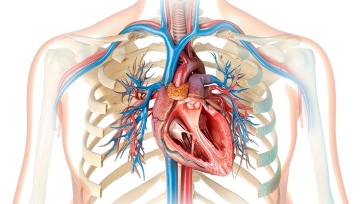
Aortic Valve Insufficiency: Symptoms, Stages, and Treatment
Girişimsel Kardiyoloji | Prof. Dr. Kadriye Kılıçkesmez » General » Aortic [...]
Read More ➜
29
Apr
Apr
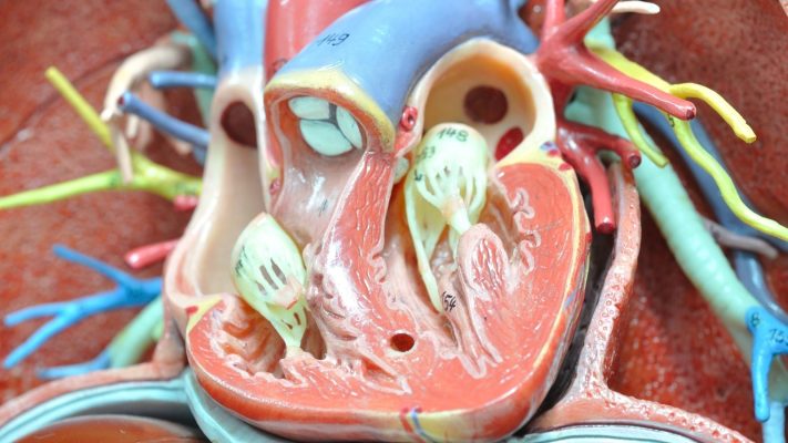
What Is Aortic Valve Regurgitation? Symptoms and Treatment
Girişimsel Kardiyoloji | Prof. Dr. Kadriye Kılıçkesmez » General » What [...]
Read More ➜
29
Apr
Apr

What Is a Heart Valve? Heart Valve Diseases, Symptoms, and Treatments
Girişimsel Kardiyoloji | Prof. Dr. Kadriye Kılıçkesmez » General » What [...]
Read More ➜
29
Apr
Apr
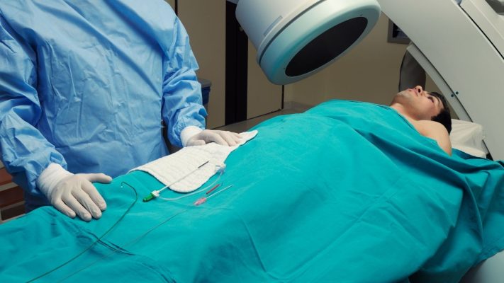
What Is Angiography? How Is It Performed and What Are the Risks?
Girişimsel Kardiyoloji | Prof. Dr. Kadriye Kılıçkesmez » General » What [...]
Read More ➜
29
Apr
Apr

What Is Rheumatic Heart Disease? Symptoms and Treatment
Girişimsel Kardiyoloji | Prof. Dr. Kadriye Kılıçkesmez » General » What [...]
Read More ➜
29
Apr
Apr

What Is Coronary Artery Disease? Symptoms, Causes, and Treatments
Girişimsel Kardiyoloji | Prof. Dr. Kadriye Kılıçkesmez » General » What [...]
Read More ➜
29
Apr
Apr
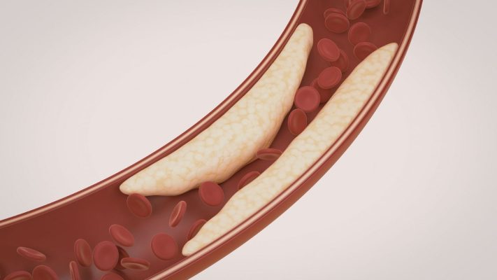
What Is Cardiovascular Blockage? Symptoms and Treatment
Girişimsel Kardiyoloji | Prof. Dr. Kadriye Kılıçkesmez » General » What [...]
Read More ➜
29
Apr
Apr

What Is Heart Failure? Symptoms and Treatment of Heart Failure
Girişimsel Kardiyoloji | Prof. Dr. Kadriye Kılıçkesmez » General » What [...]
Read More ➜
29
Apr
Apr