Türkçe
 Français
 Italiano
The prominent symptoms of a heart attack include a severe feeling of pressure in the center of the chest, pain radiating to the left arm or jaw, shortness of breath, and cold sweating. The primary cause of a heart attack is the gradual hardening of the coronary arteries that supply the heart, followed by their complete blockage by a sudden blood clot, leaving the heart muscle without oxygen. Heart attack treatment is based on mechanically opening the blocked vessel very quickly with a balloon and stent through an angiography procedure performed in hospital catheterization laboratories. In this condition, where every minute matters, immediately restoring blood flow prevents permanent tissue damage and saves lives.
| Most common symptoms | A feeling of pressure, tightness, burning, or crushing in the center of the chest; pain radiating to the left arm, shoulder, back, neck, or jaw; shortness of breath; cold sweating; nausea; vomiting; dizziness; feeling faint; extreme weakness. |
| Symptoms in women | Chest pain may be milder in women. More atypical symptoms such as shortness of breath, extreme fatigue, stomach discomfort, back or jaw pain, nausea, and dizziness may occur. |
| Symptoms in older adults | Symptoms may be less obvious in older individuals. Sudden weakness, confusion, shortness of breath, or fainting may occur as the only symptom. |
| Symptoms in patients with diabetes | Chest pain may not be felt due to diabetic neuropathy. In what is called a silent heart attack, shortness of breath, weakness, or sweating may be more prominent. |
| Causes of a heart attack | The most common cause is the rupture of atherosclerotic plaques in the coronary arteries and the formation of a clot on them. Less commonly, vessel spasm, coronary embolism, or vessel rupture may also be a cause. |
| Risk factors | Smoking, high blood pressure, high cholesterol, diabetes, obesity, a sedentary lifestyle, unhealthy nutrition, advanced age, male sex, a family history of heart disease at an early age, chronic stress, and excessive alcohol use. |
| What should be done during a heart attack? | 112 should be called immediately. The person should be allowed to rest, kept in a sitting position if possible, and should not try to go to the hospital on their own. If loss of consciousness occurs, CPR should be performed by people trained in basic life support. |
| Diagnostic methods in the hospital | The diagnosis is confirmed with electrocardiography (ECG), cardiac enzymes in the blood such as troponin, physical examination, echocardiography, and coronary angiography when necessary. |
| Emergency treatment | The aim is to open the blocked vessel as quickly as possible. Primary percutaneous coronary intervention (angioplasty and stent) is preferred together with medication treatments. Clot-dissolving drugs may be used in unsuitable cases. |
| Medication treatment | Antiplatelet drugs, anticoagulants, statins, beta blockers, ACE inhibitors or ARBs, and nitrates when necessary are the main components of treatment. Treatment is tailored to the individual. |
| Interventional treatment | The blocked vessel can be opened using a balloon and stent with coronary angiography. Coronary bypass surgery may be necessary in some patients. |
| Recovery process | The patient’s condition depends on the degree of damage to the heart muscle and the treatment applied. Regular medication use, cardiac rehabilitation, and lifestyle changes support recovery. |
| Possible complications | Serious complications such as heart failure, rhythm disorders, cardiogenic shock, heart muscle rupture, another heart attack, and sudden cardiac arrest may develop. |
| Prevention methods | Quitting smoking, regular exercise, healthy nutrition, maintaining an ideal weight, keeping blood pressure, cholesterol, and diabetes under control, regular health checkups, and regular use of prescribed medications reduce the risk. |
| When should emergency help be sought? | If there is chest pain or pressure lasting longer than 5 minutes, or symptoms such as shortness of breath, cold sweating, fainting, or pain radiating to the jaw or arm accompanying chest pain, 112 should be called without delay and emergency medical assistance should be requested. |
| Prognosis | Early diagnosis and rapid treatment significantly increase the chance of survival. Long-term outcomes vary depending on the degree of vascular damage, adherence to treatment, and control of risk factors. |
| Follow-up | Regular cardiology checkups after a heart attack, uninterrupted use of medications, adherence to an exercise program, and continuation of lifestyle changes are of great importance for long-term heart health. |
How Does a Heart Attack Begin in Our Body and What Are the Causes of Vascular Blockage?
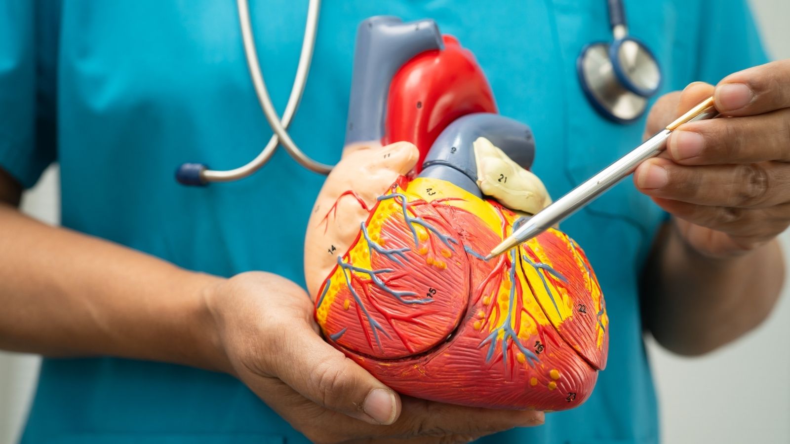
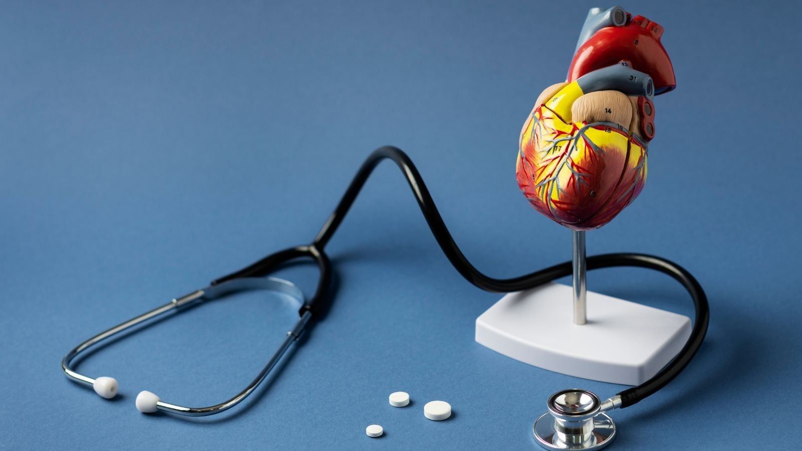
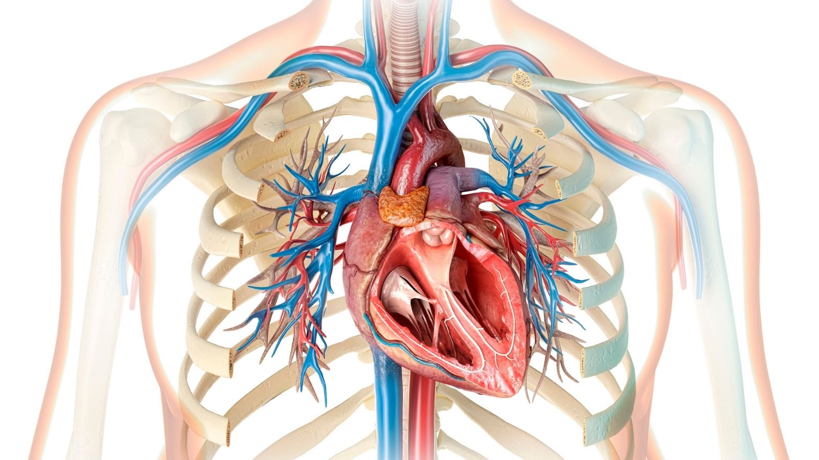
The heart is like an extraordinary engine that pumps blood, the fluid of life, to every part of the body. However, for this powerful engine to work continuously, it also needs its own fuel, namely clean blood rich in oxygen. These special vessels that surround the heart muscle like a network and nourish it are called coronary arteries. Acute coronary syndromes occur when the blood flow passing through these vessels is suddenly interrupted.
The main underlying cause of this process is atherosclerosis, commonly known as hardening of the arteries. Over the course of a silent process lasting years, cholesterol, calcium, and various inflammatory cells circulating in the blood attach to the inner walls of the coronary vessels and gradually accumulate. These deposits are called plaques. It is possible to compare this condition to the inner surface of a water pipe used for years gradually becoming narrowed by limescale. However, the event that actually triggers a heart attack is not this narrowing alone.
The outer surface of these cholesterol-filled plaques in the vessel wall may sometimes suddenly crack, rupture, or erode. When the plaque suddenly ruptures, the natural fluid structure of the blood inside the vessel is disrupted. The body’s defense and repair mechanisms perceive this rupture as a bleeding wound and immediately try to repair it. Platelets in the blood, which are responsible for clotting, rapidly rush to this area. Within seconds, a very powerful clotting cascade is triggered. Unfortunately, this healing effort by the body backfires, and the large clot mass that forms completely blocks the already narrowed vessel and abruptly stops blood flow.
When the vessel becomes blocked and blood flow is interrupted, the heart muscle cells in the area supplied by that vessel are deprived of oxygen and enter a state of illness. If this blockage is not treated and the vessel is not opened within minutes or, at the latest, a few hours, these cells begin to die permanently. When irreversible cell death occurs, the heart muscle suffers permanent damage and loses its ability to contract.
What Are the Risk Factors That Damage Vascular Structure and Are Considered Among the Causes of a Heart Attack?
There are many factors that initiate the formation of plaques inside the vessel, accelerate their growth, and create the conditions for their sudden rupture. When these factors, known as traditional risk factors, are not brought under control, the vessels undergo a continuous process of wear and tear.
The main risk factors that accelerate this process are as follows:
- High blood pressure
- High cholesterol
- Diabetes
- Smoking
- Obesity
- Sedentary lifestyle
- Genetic predisposition
- Advanced age
High blood pressure, namely hypertension, continuously damages the inner vessel wall just as pressurized water erodes a pipe, creating a suitable environment for plaque formation. High levels of what is known as bad cholesterol are the main building block of fat accumulation in the vessel wall and directly fill the plaques.
Diabetes directly damages the inner structure of the vessels due to high blood sugar levels in the circulation. It damages the structure of the entire vascular system. Tobacco use, in addition to causing sudden narrowing of the vessels, introduces toxic chemicals into the body that increase the blood’s tendency to clot, causing plaques to rupture much more easily and clots to form more readily.
Not only these well-known factors, but sometimes unexpected mechanisms can also block the vessels. Sudden severe constriction of the coronary vessels, spontaneous rupture of the vessel wall without an external cause, or a clot breaking off from another part of the body and reaching the heart may cause a heart attack. Although heart attacks have increased among young people in recent years, increasing age also emerges as a risk factor.
What Are the Symptoms of a Heart Attack That Require Emergency Intervention?
The success of a timely medical intervention depends on the signs given by the body during the heart attack being correctly interpreted by the patient and those around them. Although the symptoms do not occur with the same intensity in every person, the body generally gives very clear warning signals.
The most common symptoms encountered during a classic heart attack are as follows:
- Severe chest pain
- Pain radiating to the left arm
- Pressure radiating to the right shoulder
- Aching pain radiating toward the jaw
- Pain in the back area
- Shortness of breath
- Cold sweating
- Nausea
- Dizziness
- Intense fear of death
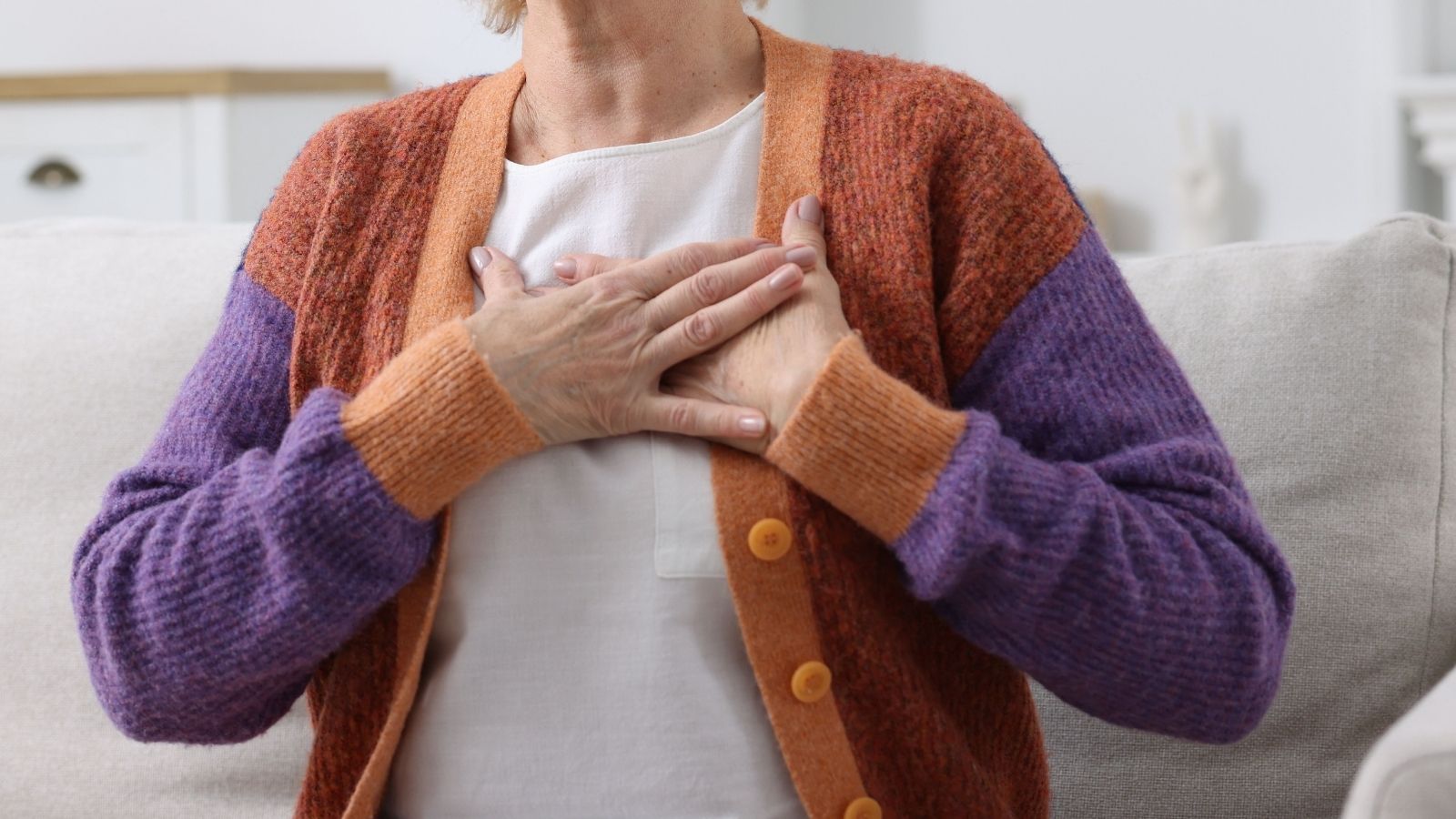
The clearest and most common symptom of acute myocardial infarction is a pressure-like pain felt in the exact center of the chest. Rather than a simple ache, this sensation is generally described as if a heavy weight has been placed on the chest, as if it is being squeezed in a vise, or as if something is being crushed inside. This relentless pain does not remain fixed only in the chest; it often spreads to the left arm, neck, lower jaw, or back. In particular, the spread of pain toward the right arm and right shoulder is one of the findings that most strongly confirms the possibility of a heart attack from a medical perspective.
Classic heart attack pain does not go away by changing your sitting position, standing up, taking a deep breath, or drinking water. It generally lasts longer than twenty minutes. Shortness of breath accompanying chest pain indicates that the heart’s pumping power has decreased and fluid has begun to accumulate in the lungs. Because the nervous system experiences intense panic during a heart attack, icy cold sweating, trembling, numbness, palpitations, and an indescribable fear of death occur in the body.
Are Heart Attack Symptoms Different in Women and People with Diabetes?
The scenario frequently seen on television or in films, in which a person collapses while clutching their chest, does not always occur in real life. Especially in women, older individuals, and patients with diabetes, the condition may develop much more insidiously with atypical symptoms.
Diabetes progresses silently over the years and damages the nerve endings in the body. This damage prevents pain signals from the heart from reaching the brain on time and correctly. Because of this neuropathy, people with diabetes may experience an infarction silently without feeling any chest pain, with only extreme fatigue, mild shortness of breath, or complaints mistaken for simple indigestion.
Women also frequently experience different signs instead of classic chest tightness. Sudden unexplained exhaustion, heartburn, a prickling sensation in the back, or unexplained cold sweating may be the only warning sign of a heart attack. Therefore, sudden-onset stomach discomfort or fatigue in patients in the risk group should be taken very seriously, and a medical evaluation should be performed without delay.
Which Tests Are Performed Before the Diagnosis and Treatment of a Heart Attack When the Patient Reaches the Hospital?
The condition of patients who present to the emergency department with suspicion is evaluated within seconds. The diagnostic process is carried out through a system that operates very rapidly according to the urgency of the patient’s condition. The first step is always to record an electrical tracing of the heart. With an ECG, disruptions in the heart’s electrical activity and the area in which the damage is located are identified within seconds.
Noninvasive imaging methods, meaning imaging methods applied from outside the body, also play a major role in diagnosis. With cardiac ultrasound, the heart’s contraction strength and regional movement abnormalities in the walls affected by the heart attack can be seen immediately on the screen. The most important finding proving that heart cells have been damaged is the cardiac enzymes measured in the blood. An increase in the enzyme called troponin in the blood is definitive evidence that heart muscle cells have begun to die.
In some uncertain cases, computed coronary angiography devices that display the anatomy of the vessels externally in three dimensions are used. If these tomography scans reveal high-risk plaques ready to rupture or severe narrowings according to a special classification system, these patients are referred without delay to catheterization laboratories, which are the main treatment centers. However, in patients who are severely overweight or have heavy calcium buildup in their vessels, these externally obtained images may not provide clear results, and it may be necessary to proceed directly to the actual angiography procedure.
How Is Heart Attack Treatment Performed Using the Angiography Method?
Catheterization laboratories, which are at the heart of interventional cardiology, are advanced technology centers where vessel-opening procedures are performed using nonsurgical methods. Percutaneous coronary interventions include all treatment methods in which the inside of the vessel is reached with very thin tubes by entering through the skin with a needle, without opening the rib cage or making large surgical incisions.
The procedures are performed while the patient is fully awake, with only the entry area numbed using local anesthesia. These procedures, which were previously performed only through the groin, are now largely performed through the vessels in the wrist because this dramatically reduces the risk of bleeding and increases patient comfort. When the procedure is performed through the wrist, the possibility of vessel damage and swelling after the procedure is much lower than with the groin.
After the area is numbed, a thin plastic sheath is placed in the vessel. Long and flexible catheters advanced through this sheath are guided along the aorta directly to the starting point of the vessels that supply the heart. A special contrast fluid visible under X-ray is injected through the catheter. X-ray devices take images from different angles to map the location and degree of the blockage and the structure of the vessel. Once imaging is complete, the treatment stage begins within seconds.
What Types of Balloons and Stents Are Used During Heart Attack Treatment?
Immediately after the diagnosis is made, physical interventions begin to open the blocked vessel. Due to the nature of interventional procedures, the instruments used are extremely precise and technologically advanced.
The main tools we use in interventional procedures are as follows:
- Guidewires
- Balloon catheters
- Drug-eluting stents
- Biodegradable stents
- Drug-coated balloons
When starting treatment, an extremely flexible guidewire is first sent into the vessel to safely cross the blockage area. This wire is gently passed through the clot or plaque. After the blockage has been crossed, a very thin, deflated balloon advanced by sliding over this wire is positioned exactly in the middle of the narrowing. This balloon is inflated with controlled pressure applied from outside. The inflated balloon crushes the hard, calcified, or clotted structure inside the vessel, compresses it toward the vessel wall, and physically widens the channel through which the blood will pass.
When only balloon dilation is performed, the risk of the vessel narrowing again due to its elastic structure is very high. Therefore, stents are used to ensure that the opened tunnel remains permanently open. A stent is a very thin wire mesh system mounted on the balloon. When it is delivered to the blocked area and the balloon is inflated, this steel or special alloy mesh opens and firmly anchors itself to the vessel wall. Acting like a tunnel support scaffold, it prevents the vessel from closing again.
Although first-generation metal stents provided structural support, the body could perceive this metal as a foreign substance and try to cover it with tissue, causing the vessel to narrow again. Drug-eluting stents, which are now used as the standard, almost completely eliminate the risk of re-narrowing by suppressing cell proliferation through the special substances on their surfaces. Stents that dissolve and disappear inside the vessel months after completing their function may also be used as a technological option in selected cases.
Which Advanced Technologies Are Used for Heart Attack Treatment in Difficult Blockages?
Not every blockage consists of a soft clot. Sometimes the vessel structure encountered is so hardened and calcified that even balloons capable of withstanding the highest pressure cannot break the calcium and widen the vessel. Much more specialized and advanced technological equipment is needed in such resistant narrowings.
In this situation, the rotablator device is used. This system has a tiny burr head coated with diamond particles at its tip. This head rotates at very high speeds inside the vessel and turns the hardened calcium blocks into dust so fine that it cannot be seen with the naked eye. After the calcium is sanded down and cleared, the vessel becomes suitable for stent placement.
Looking only from outside with X-ray does not always provide sufficient detail. It is vitally important to see whether the stent fits the vessel wall perfectly down to the millimeter and whether any tear remains at its edges. To achieve this precision, miniature cameras using sound waves or light reflections are sent into the vessel, and the inside of the vessel is scanned millimeter by millimeter. These intravascular imaging technologies greatly increase the long-term success of the procedure.
Why Is Time Vitally Important During Heart Attack Treatment?
Every second that a vessel remains blocked means that heart muscle cells deprived of oxygen are rapidly dying. The most fundamental principle used in medicine to describe this situation is the fact that time is directly equal to heart muscle. Great importance is attached to the time that passes from the moment the patient reaches the hospital until the vessel is opened with a balloon.
In major heart attacks involving complete blockage, international standards require the vessel to be opened within no more than ninety minutes from the patient’s arrival at the emergency department. If the patient presents to a smaller healthcare facility without angiography capability and will be transferred to a fully equipped center, the goal is for the total time, including the transfer process, not to exceed one hundred twenty minutes.
In scenarios where geographical conditions are very difficult and reaching angiography centers would take hours, powerful clot-dissolving drugs are used. These drugs should be administered to the patient within less than thirty minutes. However, because these drugs do not always provide complete success, the main gold standard today is to perform an ECG and make the diagnosis while the patient is on the way, then take the patient directly to the catheterization laboratory and mechanically open the vessel with a stent.
In Which Situations Is Open Heart Surgery Preferred as Heart Attack Treatment?
When the patient undergoes angiography, the picture encountered may sometimes be much more complex than the blockage of a single vessel. More than one vessel supplying the heart may be blocked or calcified along its entire length, or the main trunk vessel of the heart may be narrowed. In such extensive damage, the decision is not made by a single physician; instead, a large heart team consisting of cardiology specialists and cardiac surgeons meets and determines the most suitable option for the patient’s anatomy.
The situations in which surgery is more commonly preferred are as follows:
- Complex three-vessel blockage
- Left main coronary artery stenosis
- Widespread damage due to diabetes
- Severely reduced heart contraction strength
- Mechanical ruptures occurring in the heart
Large-scale scientific studies have proven that performing coronary bypass surgery instead of placing stents provides much more successful long-term results in patients with very complex vascular anatomy, especially those with insulin-dependent diabetes. Diabetes damages the vessels so extensively that placing a stent in every centimeter of the vessel is neither possible nor healthy.
Likewise, if a rupture has occurred in the wall or valves of the heart during a heart attack, surgical intervention becomes unavoidable. In many patients who do not have these complex conditions or urgent mechanical damage, stenting procedures are a powerful alternative that protects the patient from major surgery, reduces the risk of infection, and speeds up the return to normal life to an extraordinary degree.
Which Medications Must Be Used After Heart Attack Treatment?
Opening the blocked vessel with a successful angiography and stent procedure is only the first and most urgent stage of the process. The patient is monitored in intensive care and discharged home after the entry site in the leg or wrist has healed. However, the actual treatment process begins outside the hospital. A highly disciplined medication routine is essential to prevent clot formation inside the stent placed in the heart and to prevent the heart attack from recurring months later.
The main groups of medications that must be used after discharge are as follows:
- Blood thinners
- Cholesterol-lowering medications
- Beta blockers
- Blood pressure regulators
- Stomach protectors
The backbone of medication treatment is the dual blood-thinner protocol. Together with aspirin, a much stronger second antiplatelet drug is generally used without interruption for at least one year. These medications prevent platelets from sticking to one another, making it impossible for the inner surface of the stent to fill with a clot.
Statin-group cholesterol-lowering medications do not merely reduce the amount of fat in the blood; more importantly, they stabilize the surfaces of plaques inside the vessels like a protective coating and prevent new ruptures. Blood pressure and rhythm regulators that reduce the workload of the heart are also added to the prescription to prevent the heart from changing shape and enlarging over time due to the damage caused by the heart attack.
Frequently Asked Questions

Prof. Dr. Kadriye Orta Kılıçkesmez is one of the leading figures in the field of Turkish cardiology. She was born on January 24, 1974, in Tekirdağ. After completing her undergraduate education at Istanbul University Cerrahpaşa Faculty of Medicine, she chose cardiology as her specialty and received her specialist training at the Cardiology Institute of the same university. In 2015, she was appointed by the university to establish the Şişli Etfal cardiology clinic and Angio laboratory. Becoming a professor in 2017, Kadriye Kılıçkesmez established the cardiology clinic and Angio laboratory of Prof. Dr. Cemil Taşçı Hospital in 2020 and ensured that the clinic became a training clinic.