Türkçe
 Français
 Italiano
The main difference between TriClip and tricuspid valve surgery is the way the intervention is performed and the recovery process. The TriClip procedure is a minimally invasive method in which a catheter is inserted directly through the groin vein without opening the chest, and a clip is attached to the leaking valve. Conventional open surgery, on the other hand, requires cutting the breastbone, stopping the heart, and using a heart-lung machine. While open surgery involves a long intensive care stay, a difficult bone-healing period, and high risk, minimally invasive clipping technology allows the patient to return home within a few days thanks to its incision-free structure. For elderly patients with weakened hearts, this innovative approach completely eliminates the heavy physical burden of conventional surgery.
Where Is the Tricuspid Valve Located in the Heart and What Is Its Function?
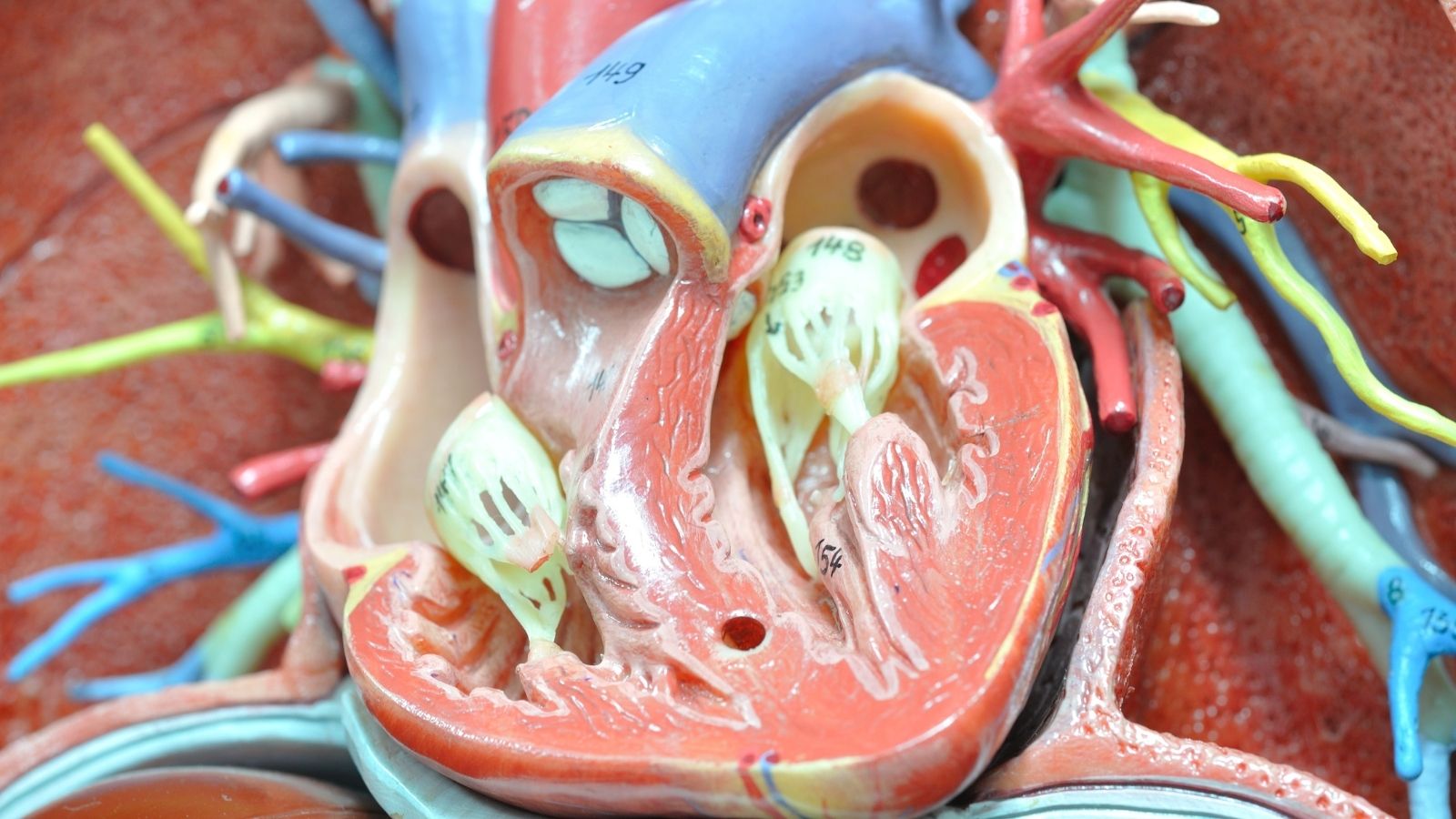
Our heart is a four-chambered pump system that distributes blood throughout the body and to the lungs. On the right side of this structure, there is a dynamic gateway between the upper chamber and the lower main pumping chamber that controls the passage of blood. This structure, called the tricuspid valve, allows oxygen-depleted blood returning to the heart to pass into the lower right chamber. When the heart contracts, it attempts to prevent this blood from flowing backward to where it came from. For blood to move in one direction toward the lungs to be oxygenated, this valve is expected to open and close regularly with every heartbeat.
For many years in the medical world, the valves on the left side of the heart received greater attention, while the tricuspid valve generally remained in the background. For this reason, it was sometimes even referred to as the forgotten valve. However, clinical observations obtained in recent years have revealed how decisive the balance of blood flow on the right side of the heart is for overall health. Under normal physiological conditions, the speed and pressure of blood passing through the tricuspid valve are based on a highly delicate balance. In this low-pressure system, deterioration in the structural integrity of the valve may cause backward pressure loading in the right chambers of the heart. Over time, this may initiate a process affecting the entire body system. The valve leaflets are attached to the lower chamber by thin cords and muscles. This complex anatomical structure must function harmoniously as a whole for the valve to work properly.
Why Do Tricuspid Valve Diseases Develop and How Do They Affect the Heart?
In clinical practice, tricuspid valve diseases mainly appear as narrowing of the valve or the inability of the valve to close completely. Tricuspid stenosis may generally be associated with the effects of rheumatic diseases experienced during childhood. This condition is relatively rare and tends to occur together with narrowing of the valves on the left side of the heart rather than appearing alone. When the valve area falls below a certain level, the passage of blood becomes difficult.
The condition encountered more frequently today is tricuspid regurgitation. Regurgitation is the inability of the leaflets forming the valve to come together and provide adequate sealing when the heart contracts. As a result, while blood should move forward, some of it leaks backward. The vast majority of tricuspid regurgitation cases develop because of other diseases rather than a disorder in the valve tissue itself. Causes such as left-sided heart problems, chronic lung diseases, or increased pressure in the pulmonary vessels may cause the lower right chamber of the heart and the frame on which the valve sits to stretch over time. As the frame widens, the valve leaflets move apart, leaving a gap in the middle and allowing blood to leak backward. This mechanism may gradually tire and enlarge the right side of the heart.
What Symptoms Occur in the Body If Tricuspid Valve Regurgitation Is Not Treated?
When this leakage on the right side of the heart is not controlled, blood accumulating in the right chamber begins to create backward pressure throughout the venous system. The vessels carrying oxygen-depleted blood from the body to the heart are directly affected by this increased pressure. Problems such as fluid accumulation in different parts of the body and blood congestion in the organs may begin to appear. Patients generally experience gradually increasing fatigue and may attribute it to aging or daily stress. However, as the disease progresses, the complaints begin to significantly restrict daily life.
The main observable symptoms are as follows:
- Prominent neck veins
- Enlargement of the liver
- Fluid accumulation in the abdomen
- Swelling in the legs
- Edema in the ankles
- Easy fatigue
- General weakness
- Loss of appetite
- Shortness of breath
When these symptoms appear, the patient’s walking distance may shorten and exercise capacity may decrease. The liver may enlarge because of exposure to high pressure and may cause tenderness beneath the right rib cage. When the fluid portion of the blood begins to leak outside the vessels, fluid accumulation called ascites may occur in the abdominal cavity. Edema in the ankles may become more noticeable later in the day because of the effect of gravity. Since the organs cannot receive the blood circulation they require completely, patients may experience severe fatigue. In untreated cases, kidney and liver functions may begin to slow in advanced stages.
How Is the Decision for Conventional Open-Heart Surgery Made and What Are Its Risks?
Planning surgical intervention for tricuspid regurgitation is considered in order to prevent permanent exhaustion of the right ventricle. Conventional surgical options include repairing or replacing the valve through methods in which the patient’s chest is opened or access is gained between the ribs. During echocardiographic examinations, the structure of the valve, the degree of regurgitation, and the size of the right heart are measured in detail. Surgical repair may be considered when the valve frame has enlarged beyond a certain level.
The patient’s general health status, age, and surgical risk are decisive in this decision. Especially if other valves on the left side of the heart will also be treated, repairing the tricuspid valve during the same session may be preferred. The general surgical approach is to repair the patient’s natural valve rather than replace it whenever possible. Preserving the natural valve may reduce the need for blood-thinning medications in the future. However, if there is extensive calcification or infection in the valve, repair becomes more difficult and replacement with a biological or mechanical prosthesis may be required.
Isolated open-heart surgeries performed only on the tricuspid valve may carry certain risks. The main factors determining this risk are that patients are generally older and that their bodies have become exhausted because of long-standing heart failure. Other organs such as the liver and kidneys may also be affected by backward blood pressure. Scientific scoring systems show that open-heart surgery may be risky for elderly patients. In such cases, minimally invasive methods stand out as an important alternative for overcoming the difficulties of open surgery.
What Is the TriClip Method in Interventional Cardiology and How Is It Performed Without Surgery?
Transcatheter edge-to-edge repair technology is a modern method that makes it possible to treat heart valves without opening the chest or stopping the heart. The TriClip system is considered an important option for patients at high risk for open surgery. This method is performed using a completely minimally invasive approach and does not require any surgical incision in the chest area.
The procedure begins by creating a very small entry point in the vein in the patient’s groin. This vascular access is used to advance toward the heart. Once the right atrium is reached, the TriClip device, which resembles a clip and is located at the tip of a special delivery system, comes into use. This technology is designed to adapt to the wide, three-leaflet anatomical structure of the tricuspid valve. One of the technical features of the device is its ability to grasp the leaflets independently from one another.
In patients with gaps between the valve leaflets, the physician may first grasp one leaflet with the device and then draw the other leaflet into the device to join them together. In this way, the opening in the center of the valve is narrowed and the amount of blood leaking backward is mechanically reduced. Since the procedure is performed while the heart continues beating, the intervention in the patient’s bodily functions is highly limited.
How Is the TriClip Procedure Performed Step by Step and What Is the Process Like?
This treatment method is performed in fully equipped catheterization laboratories. To ensure patient comfort and allow the valve to be evaluated clearly, the procedure is generally carried out under general anesthesia or deep sedation. The heart is not stopped as it is in open surgery; it continues pumping blood. After reaching the heart through the groin vein, physicians navigate using continuous X-ray imaging and three-dimensional echocardiography devices that display the heart in detail through the esophagus.
The main steps during the procedure are as follows:
- Establishing vascular access through the groin
- Advancing the catheter
- Three-dimensional imaging
- Evaluating the valve
- Positioning the clipping system
- Checking the leakage
- Removing the catheter from the body
With the guidance of images obtained through the esophagus, the clip is directed toward the leaking area of the valve. When the valve leaflets are grasped and the device is closed, the aim is to reduce the amount of blood leaking backward. One of the advantages of this procedure is that the effect of the intervention can be observed on the screen within seconds. If the leakage is not reduced sufficiently, the clip can be reopened and repositioned, or a second clip may be added depending on the size of the valve. The procedure is generally completed within a few hours, and the device placed on the valve gradually becomes covered with tissue and remains in place as though it were a natural part of the heart.
What Are the Differences Between TriClip and Open-Heart Surgery in Terms of Incisions and Intensive Care?
The differences between the two treatment approaches directly affect the comfort of the recovery process. Conventional tricuspid valve surgery generally requires opening the breastbone. To repair the heart, the patient’s heart is stopped and vital functions are transferred to a machine that circulates blood outside the body. This process may cause various reactions in the body.
In the TriClip method, the breastbone is not touched. Only a vascular entry through the groin is used, and this small entry site is closed when the procedure is completed. Repair is performed without the need for a heart-lung machine while the organs continue functioning in their natural rhythm. This may make the recovery process easier, especially for elderly patients or those with reduced lung capacity.
The main differences in the post-treatment period are as follows:
- Length of hospital stay
- Need for intensive care
- Healing of the incision site
- Speed of returning to normal life
- Duration of physical restrictions
After open surgery, patients may remain in intensive care for longer periods and their discharge time may be prolonged. After discharge, healing of the breastbone may take weeks. During this period, it is important for patients to avoid lying on their side or lifting heavy objects. In the TriClip method, intensive care monitoring is generally brief and only precautionary. Most patients can be discharged within a few days. Since there is no chest incision, bone healing is not required, and patients can return to their daily activities in a short time. Only minor physical restrictions may need to be observed during the first few days to allow the groin area to heal.
What Are the Differences Between TriClip and Surgery in Terms of Pacemaker Risk and Medication Use?
The position of the tricuspid valve in the heart is very close to the heart’s natural electrical system. During open surgery, especially when the valve is replaced, the procedures performed may affect these electrical conduction pathways. In such cases, a pacemaker may need to be implanted after the procedure. In the TriClip procedure, since no sutures are placed in the valve tissue, the likelihood of pressure on the electrical pathways is much lower. Therefore, the need for a permanent pacemaker is also relatively rare.
There are also differences in terms of the medications used. If a mechanical valve is implanted through open surgery, patients may need to use very strong blood-thinning medications and undergo frequent blood tests to prevent clot formation. This period may be shorter with biological valves, but the lifespan of the prosthesis may be limited. Since the patient’s own valve tissue is preserved in the TriClip procedure, very intensive blood-thinning regimens are generally not required unless the patient has another underlying rhythm disorder. The process can usually be managed with more commonly used medications suitable for the patient’s general condition. This places fewer restrictions on the patient’s daily life and diet.
What Are the Success Rates of TriClip Treatment in Light of Current Data from Türkiye?
Transcatheter repair technology is also applied in our country and closely monitored clinically. National clinical studies in the literature have examined patient groups whose symptoms continued despite medication and who were considered to have a very high surgical risk. The results of this minimally invasive method are promising in elderly individuals with accompanying diseases who are considered unsuitable for open surgery.
Procedural success rates in placing the device in the target area are high. In a significant proportion of patients, the amount of blood leaking backward through the valve showed a marked tendency to decrease compared with the pre-procedure period. Reducing the valve leakage relieves the burden on the right side of the heart and contributes to improvement in the patient’s symptoms. During follow-up periods, reductions have been recorded in the rates of rehospitalization because of heart failure.
Improvements may be observed in quality-of-life scales and walking tests. The fact that the procedural risk remains at more reasonable levels compared with open surgery supports the acceptance of this method as a safe alternative for elderly patients. As with every medical intervention, the results may vary according to the condition of the patient’s heart muscle and accompanying diseases. However, the overall picture indicates that the hemodynamic benefit provided by the procedure may improve quality of life.
At What Stage Should Patients with Heart Failure Be Evaluated for TriClip Treatment?
Tricuspid valve regurgitation is a disease that may progress gradually over time. Seeking medical attention may be delayed because symptoms such as fluid retention, weakness, or shortness of breath are thought to be related only to aging. If patients no longer obtain the expected benefit from the diuretic medications they use or if swelling in the legs has begun to restrict daily activities, they may need to be evaluated for minimally invasive treatment options. Intervening before the heart muscle reaches an irreversible level of exhaustion may increase the chance of success.
During this evaluation process, patients are examined by a multidisciplinary Heart Team consisting of cardiologists, cardiac surgeons, and imaging specialists. The patient’s echocardiographic findings, pulmonary pressure, and functions of other organs are evaluated together.
The main steps in the evaluation process are as follows:
- Echocardiographic examination
- Analysis of right-heart functions
- Measurement of pulmonary pressure
- Assessment of kidney and liver functions
- Calculation of surgical risk scores
- Review of accompanying diseases
If the patient is elderly, has lung disease, and open-heart surgery is considered physically demanding, the Heart Team may direct the patient toward interventional treatments performed through the groin. This decision-making process, applied to the right patient at the right time, directly affects the patient’s response to treatment.
What Should Patients Pay Attention to After Minimally Invasive TriClip Treatment?
The recovery process after the procedure generally progresses very quickly. Although returning to daily life after leaving the hospital is easy, it is important to continue certain lifestyle adjustments because heart failure is an ongoing process. Although the intervention on the valve provides mechanical correction, patients still have certain responsibilities to help reduce the burden on the heart.
The points that should be considered after discharge are as follows:
- Avoiding strain on the groin area
- Avoiding heavy lifting during the first few days
- Using the recommended medications regularly
- Restricting salt consumption
- Monitoring daily fluid intake
- Beginning regular walks
- Attending follow-up appointments
Restricting salt intake is highly important for preventing the body from retaining fluid again. Even if the valve leakage decreases, the recovery period of the heart muscle continues. Not stopping the medication treatment recommended by physicians suddenly and not neglecting echocardiographic follow-up appointments support the recovery process.
Frequently Asked Questions
TriClip is an interventional method performed through the groin and does not require open-heart surgery. Tricuspid valve surgery involves surgically repairing or replacing the valve. The choice of treatment is determined according to the individual patient.
Hospital stay and recovery time are generally shorter after TriClip. In open-heart surgery, recovery may take longer because the breastbone needs to heal. The duration may vary according to the patient’s general health condition.
TriClip is a minimally invasive treatment option considered especially in suitable patients with severe tricuspid valve regurgitation who are elderly or at high risk for open-heart surgery.
If there is advanced structural damage to the valve, damage that cannot be repaired, or anatomical features unsuitable for TriClip, surgical valve repair or valve replacement may be preferred.
The severity of valve regurgitation, the pumping strength of the heart, the patient’s age, accompanying diseases, and general health status before the procedure are among the main factors affecting treatment success.
Most patients can get up within a short time and return to daily activities within a few days. However, the timing of returning to physical activity should be planned according to the patient’s clinical condition and physician recommendations.
After both treatments, valve functions are evaluated through echocardiography and cardiology checkups. Regular follow-up helps detect possible complications early and monitor the success of the treatment.
Medication treatment is determined according to the patient’s rhythm disorder, valve condition, and other diseases. Blood-thinning medications or other heart medications should be used only as recommended by the cardiologist.
After successful treatment, symptoms such as shortness of breath, weakness, and swelling in the legs may decrease. As a result, exercise capacity and daily quality of life may improve significantly in many patients.
The treatment decision is made individually by the Heart Team after evaluating the valve structure, severity of regurgitation, the patient’s age, surgical risk, and accompanying diseases together.

Prof. Dr. Kadriye Orta Kılıçkesmez is one of the leading figures in the field of Turkish cardiology. She was born on January 24, 1974, in Tekirdağ. After completing her undergraduate education at Istanbul University Cerrahpaşa Faculty of Medicine, she chose cardiology as her specialty and received her specialist training at the Cardiology Institute of the same university. In 2015, she was appointed by the university to establish the Şişli Etfal cardiology clinic and Angio laboratory. Becoming a professor in 2017, Kadriye Kılıçkesmez established the cardiology clinic and Angio laboratory of Prof. Dr. Cemil Taşçı Hospital in 2020 and ensured that the clinic became a training clinic.