Türkçe
 Français
 Deutsch
 Italiano
 Español
 العربية
 Русский

LAA closure (left atrial appendage closure) is a nonsurgical interventional cardiology procedure performed to eliminate the risk of stroke in patients with heart rhythm disorders caused by atrial fibrillation. With this method, the small pouch in the left atrium, considered the main source of blood clot formation in the heart, is permanently isolated using a special device in an angiography laboratory.
The standard approach to preventing blood clot formation associated with heart rhythm disorders is the use of blood-thinning medications. However, lifelong use of these medications becomes unsustainable for individuals who have a high risk of bleeding or have previously experienced serious bleeding episodes.
The left atrial appendage closure procedure completely blocks the passage of blood clots into the systemic circulation through a mechanical barrier. Thus, patients receive safe protection against the risk of stroke without remaining dependent on medications that carry a high risk of bleeding.
What Is This Pouch That Creates the Need for LAA Closure (Left Atrial Appendage Closure)?
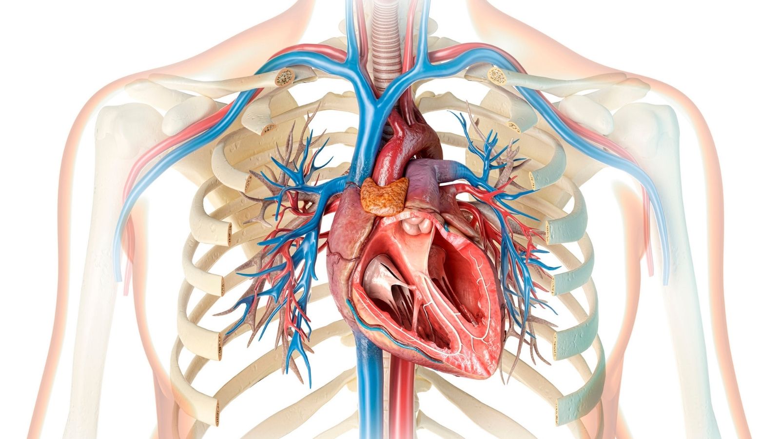
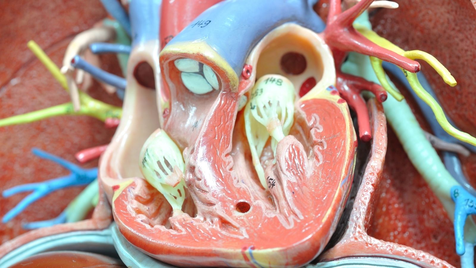
The heart is a highly complex organ with its chambers, valves, and various internal structures. Certain special structures inherited from our developmental period in the womb remain part of our hearts throughout our lives. The left atrial appendage is a small pouch located in the upper left chamber of the heart (left atrium), characterized by its elongated, irregular shape and closed tip. Its shape can be compared to the appendix in our intestines, although its function is quite different. Rather than having a smooth inner wall, the inner surface of this pouch has a mesh-like, ridged structure composed of small muscle bundles.
In a normal and healthy heart rhythm, this pouch performs a highly beneficial function. When a large amount of blood enters the upper chamber of the heart and pressure increases, its flexible structure allows it to expand and function as a type of pressure-balancing chamber. Every time the heart contracts, this pouch also contracts forcefully in coordination with the main rhythm, emptying the blood inside it into the main chamber. In the presence of a healthy rhythm, this pouch fills with fresh blood and empties with every heartbeat. Therefore, blood does not remain inside or become stagnant. However, when the heart’s natural rhythm is disrupted, this small and seemingly harmless structure can become a suitable environment for blood clot formation.
Why Does LAA Closure (Left Atrial Appendage Closure) Become Important in Heart Rhythm Disorders?
Atrial fibrillation is one of the most common heart rhythm disorders in the population, particularly with advancing age. In this condition, the upper chambers of the heart lose their ability to contract regularly and in coordination. Instead of strong pumping movements, these chambers begin to experience very rapid and irregular vibrations. This disruption in the heart’s main rhythm directly affects blood flow within the left atrial appendage.
We can compare this to a stream in nature. In a river that flows rapidly and regularly, the water is constantly moving and does not accumulate algae. However, if a motionless, stagnant inlet forms along the riverbank, disconnected from the main current, sediment begins to accumulate there. When a heart rhythm disorder develops, the left atrial appendage similarly becomes a stagnant inlet within the heart. Because regular contractions are lost, blood flow inside the pouch slows down significantly.
Blood is naturally a fluid that must remain in continuous motion. When movement slows and blood cells become stagnant between those irregular walls, the body’s clotting mechanisms may become activated unintentionally. Observations show that, in heart rhythm disorders unrelated to heart valve disease, the vast majority of blood clots forming in the heart tend to accumulate inside this pouch. The possibility that these clots may break away and spread throughout the body is the main factor that leads us to take more protective measures.
How Is the Risk of Blood Clots Managed Before LAA Closure (Left Atrial Appendage Closure)?
A blood clot that forms inside this pouch generally does not cause discomfort or symptoms as long as it remains in place. The problem begins when the clot breaks away from its location and enters the bloodstream. A clot that becomes dislodged as a result of a sudden change in blood pressure or a fluctuation in heart rhythm enters the heart’s main pumping chamber and is then pumped into the body through the aorta. Since the blood vessels supplying the brain are among the first destinations of blood leaving the heart, the risk of the clot blocking a blood vessel in the brain is quite high. This condition is called ischemic stroke.
In medicine, the primary method we have used for many years to manage this risk is anticoagulant medication, commonly known as blood thinners. These medications work by reducing the blood’s tendency to clot throughout the body and provide substantial protection against the risk of stroke. However, every treatment has its own challenges. When the blood’s ability to clot is weakened, the body’s natural ability to stop ordinary bleeding also slows down.
For patients, this situation often requires a delicate balance. When the medication dose is slightly too high, bleeding risks may arise in the stomach, intestines, or the brain, which is a much more sensitive area. When the dose is insufficient, protection decreases and the risk of clot formation reappears. Particularly in individuals above a certain age, those with accompanying medical conditions, or those prone to injury, long-term use of these medications may become unsafe. In such cases, taking a physical preventive measure directed at the source of the problem becomes a more reasonable option.
Who Is Considered a Suitable Candidate for LAA Closure (Left Atrial Appendage Closure)?
This procedure is not a treatment considered as the first step for every patient with a heart rhythm disorder. It is primarily planned for individuals who are considered to have a high risk of stroke but for whom taking blood-thinning medications poses significant risks to their health. The patient’s condition is evaluated from multiple perspectives, and this procedure is considered when the potential harm of medication outweighs its benefits.
Patient groups who may be candidates include:
- Individuals with a history of stomach bleeding
- Individuals who have experienced intestinal bleeding
- Individuals who have survived a brain hemorrhage
- Elderly individuals at high risk of falling
- Patients with advanced kidney disease
- Individuals who develop blood clots despite medication
When we examine these groups more closely, we can see how delicate the situation is. For example, restarting blood-thinning medication in someone who has previously experienced a brain hemorrhage is a highly concerning situation. Similarly, in elderly patients who frequently lose their balance and fall for neurological reasons, even a minor impact can result in serious internal bleeding while taking blood thinners. In some cases, blood clot formation cannot be prevented even though patients take their medications very regularly and without missing any doses. For these patient profiles, mechanically closing the source of the problem in the heart, rather than altering blood chemistry, aims to improve quality of life and safety.
How Is the Heart’s Structure Examined Before LAA Closure (Left Atrial Appendage Closure)?
Just as human faces differ from one another, the structure of this pouch inside the heart also varies considerably from person to person. For the closure procedure to achieve the desired result, this area must be mapped in great detail before the intervention. For this purpose, advanced imaging methods such as detailed heart ultrasound performed through the esophagus (TEE) and three-dimensional cardiac tomography are generally used. During these examinations, the depth of the pouch, the width of its opening, its angles, and whether an active blood clot is present inside it at that moment are evaluated.
In medical literature, these pouches are divided into several basic types according to their shapes. These anatomical shapes are as follows:
- Chicken wing shape
- Windsock shape
- Cactus shape
- Cauliflower shape
Each anatomical structure has its own characteristics. For example, the chicken wing shape has a distinct bend after the entrance and requires calculation of the depth at which the device will be positioned. The windsock shape is straighter and deeper. The cactus and cauliflower shapes consist of numerous small lobes, and their openings are generally wider. Your doctor determines which anatomy you have by examining your imaging results and plans the model and exact size of the device to be used before the procedure based on these findings.
How Is Nonsurgical LAA Closure (Left Atrial Appendage Closure) Performed?
This procedure is not open-heart surgery requiring the chest to be opened or the heart to be stopped. It is a minimally invasive procedure performed in hospital angiography laboratories under the guidance of advanced imaging technologies. Due to the precision of the procedure and to ensure the patient’s comfort throughout, sedation that produces a deep sleep-like state or general anesthesia is generally preferred. This way, the patient does not experience pain, anxiety, or discomfort during the procedure.
The procedure begins with a very small needle puncture into a large vein in the right groin area. Guiding tubes (catheters) are then inserted into the blood vessel through this entry point. Following the blood vessel pathway, the right upper chamber of the heart is reached first. Since the target is on the left side, a special technique is used to cross the thin membrane separating the right and left chambers of the heart. This step is called transseptal puncture.
After reaching the left chamber, the catheter is carefully directed toward the opening of the left atrial appendage. The closure device, selected according to the measurements determined before the procedure, is advanced through the catheter in a compressed form. When the device reaches the opening of the pouch, it is slowly released. Thanks to its shape-memory metal structure, the device expands and covers the opening of the pouch, much like an umbrella or a flexible plug. This stage is not rushed, and various tests are performed to ensure that the device is positioned correctly.
Which Safety Criteria Are Monitored During LAA Closure (Left Atrial Appendage Closure)?
Placing the device is only one part of the procedure; what truly matters is ensuring that the device will remain there securely and permanently. Therefore, a series of standard safety checks is carefully performed before the connecting wire holding the device is released. These checks, carried out using simultaneous ultrasound and X-ray images, support the successful achievement of the procedure’s objective.
The main criteria evaluated during the procedure include:
- Correct positioning
- Attachment to tissue
- Device flexibility
- Leak prevention
To explain these steps in more detail, the first check determines whether the device is centered over the opening of the pouch and does not come into contact with neighboring valves or blood vessels. In the second step, the catheter is gently pulled backward to perform a stability test and determine whether the microscopic anchors along the edges of the device are sufficiently attached to the heart tissue. Third, the pressure exerted by the device on the heart wall and the degree of compression are measured, ensuring that it is neither too loose nor so tight that it places strain on the heart. Finally, color Doppler ultrasound is used to check whether blood is leaking around the edges of the device into the pouch. If all these criteria are satisfactory, the device is detached from the catheter, the procedure is completed, and the entry site in the groin is closed.
What Happens During Recovery After Left Atrial Appendage Closure (LAA Closure)?
After the closure procedure is completed, the patient is taken to an observation room, and the awakening process is monitored. Since open surgery is not performed, there is no long and painful recovery period associated with healing of the breastbone. The primary expectation is that the patient rests on their back for several hours without bending their leg, allowing the entry site in the groin to heal safely.
After this resting period, patients can generally stand up, walk, and eat their normal meals. The following morning, a brief ultrasound examination is performed to confirm that everything has proceeded as planned. If the evaluation reveals no obstacles, patients are generally discharged from the hospital walking on their own one day after the procedure. Following a short rest period of a few days, patients can return relatively quickly to daily activities that do not require strenuous physical exertion.
As the device remains in the body, the heart’s own natural healing mechanism begins to work. Although the device initially represents a foreign surface to the body, the cells lining the inner surface of the heart begin covering it within weeks. Over time, the portion of the device that comes into contact with blood becomes completely covered by the patient’s own natural tissue. Thus, the device becomes part of the heart, and the high-risk pouch is structurally isolated from the circulatory system.
How Is Medication Use Adjusted After LAA Closure (Left Atrial Appendage Closure)?
Although the main objective of the procedure is to discontinue strong blood-thinning medications, this does not happen immediately at the moment the procedure is performed. It takes a certain amount of time for the device to become covered by the heart’s own cells. During this period, a temporary and stepwise medication plan is implemented to prevent clot formation on the metal surface of the device.
During the first month and a half after the procedure, a relatively more protective medication regimen is administered, adjusted according to the patient’s existing bleeding risk. Approximately 45 days later, the patient is called for a follow-up examination, and a detailed ultrasound is used to assess whether the device has become covered with tissue. If the cells have sufficiently covered the device and the seal remains intact, the goal is to discontinue the use of strong blood thinners. As the sixth month approaches, the treatment plan is generally simplified to continue with only a low-dose preventive medication (most commonly a single aspirin). Thus, the days of intensive medication use associated with fear of bleeding are largely left behind.
What Are the Possible Risks of Left Atrial Appendage Closure (LAA Closure)?
Every medical intervention, regardless of how advanced it may be, carries certain inherent risks. However, thanks to current technology, experienced teams, and detailed planning before the procedure, the risks of these interventional procedures have been reduced to very low levels. Every precaution is prepared to address possible complications during the procedure.
Some possible risks include:
- Fluid accumulation in the pericardium
- Device displacement
- Bleeding in the groin
- Bruising in the groin
Because heart tissue is extremely delicate, minor fluid leakage toward the pericardium may occasionally occur as the catheter advances, although this is rare. Since continuous ultrasound monitoring is performed throughout the procedure, this situation is detected immediately and is generally controlled by draining the fluid with a small needle intervention. The possibility of the device failing to attach securely and moving out of position is also very rare thanks to the safety tests mentioned above; however, if it occurs, retrieving the device with a catheter is often possible. The most common occurrence, as with any angiography procedure, is mild bruising and swelling at the entry site in the groin, which tend to resolve spontaneously within a few days.
What Should Patients Pay Attention to After LAA Closure (Left Atrial Appendage Closure)?
After a successful closure procedure, patients return to their normal social and physical lives following a short period of rest. Since the implanted device is made from a special titanium-containing alloy, it does not trigger alarms at airport security gates or detectors in shopping centers. In addition, if patients need magnetic resonance imaging (MRI) in the future for orthopedic or neurological reasons, the vast majority of these devices are MRI-compatible and therefore do not pose a problem.
However, there is one very important detail that must not be forgotten. This procedure is not a method for treating the patient’s atrial fibrillation, namely the heart rhythm disorder itself. Irregularities in the heart’s rhythm may continue; the sole purpose of the procedure is to minimize the risk of stroke that these irregularities may cause. Therefore, it is extremely important for patients to continue taking medications that regulate their heart rate, blood pressure treatments, and other cardiovascular medications as recommended by their doctors.

Prof. Dr. Kadriye Orta Kılıçkesmez is one of the leading figures in the field of Turkish cardiology. She was born on January 24, 1974, in Tekirdağ. After completing her undergraduate education at Istanbul University Cerrahpaşa Faculty of Medicine, she chose cardiology as her specialty and received her specialist training at the Cardiology Institute of the same university. In 2015, she was appointed by the university to establish the Şişli Etfal cardiology clinic and Angio laboratory. Becoming a professor in 2017, Kadriye Kılıçkesmez established the cardiology clinic and Angio laboratory of Prof. Dr. Cemil Taşçı Hospital in 2020 and ensured that the clinic became a training clinic.