Türkçe
 Français
 Deutsch
 Italiano
 Español
 العربية
 Русский

Aortic valve replacement (AVR) is a procedure in which the aortic valve, which allows oxygenated blood to be pumped from the heart to the body but has lost its function due to calcification or insufficiency, is replaced with a prosthetic valve.
This procedure is performed using two main methods, depending on the patient’s anatomical structure and current health profile. The first option is open-heart surgery, in which the chest is opened, the damaged valve is removed, and a new valve is sutured into its place.
The other method is the minimally invasive (TAVI) approach, in which the new valve is placed in the heart by advancing thin catheters through a blood vessel in the groin without cutting the breastbone. Both techniques focus on reducing the burden on the heart and restoring healthy blood circulation.
Where Is the Aortic Valve Located in Our Heart and How Does Aortic Valve Disease Begin?
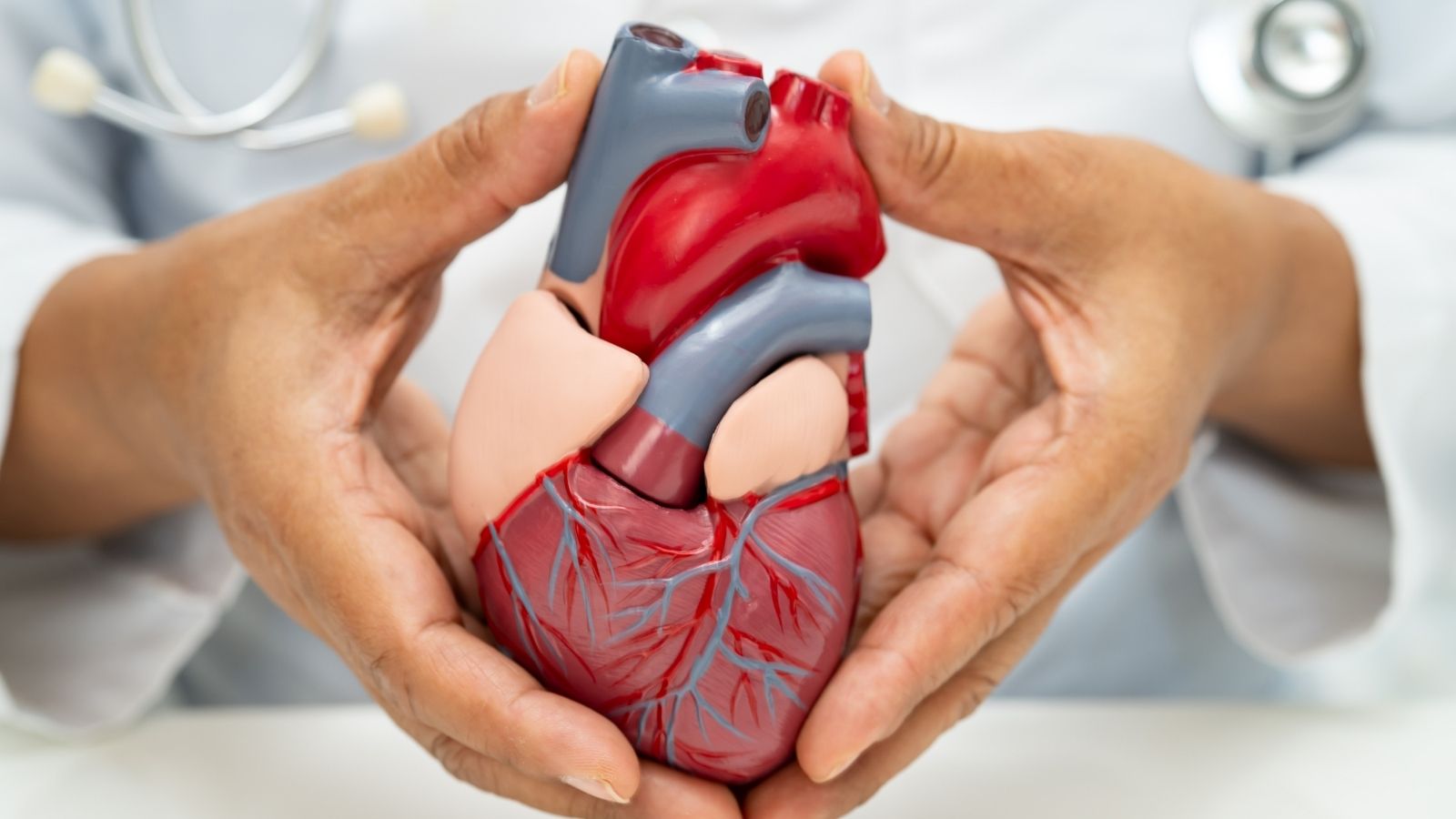
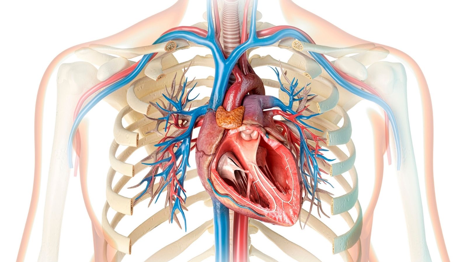
Our heart is like a continuously operating pump responsible for delivering oxygenated blood to every part of the body. The aortic valve is located at the junction between the left ventricle, the main chamber of this powerful pump, and the aorta, the largest artery in the body. Under normal circumstances, this structure, consisting of three thin and flexible leaflets, functions as a one-way safety gate. When the heart contracts, the leaflets open to allow blood to be ejected into the artery, and when the heart relaxes, they close to prevent blood from flowing backward. This mechanism, which opens and closes approximately one hundred thousand times a day, may undergo natural wear and tear over the years. With advancing age, microscopic deterioration in the tissues creates conditions that favor calcium accumulation in the area. Sometimes, having a congenital two-leaflet (bicuspid) valve can alter the distribution of stress on the valve, causing calcification to begin at an earlier age. Over time, the calcified leaflets thicken, harden, and lose their flexibility.
How Do Aortic Valve Calcification (Stenosis) and Aortic Valve Regurgitation Affect Our Body?
Calcification and narrowing of the aortic valve create a narrow passage in front of the heart. The heart must generate much higher pressure than normal to push the blood needed by the body through this restricted opening. To adapt to this increased workload, the wall of the left ventricle gradually begins to thicken, much like a muscle undergoing weight training. Although this thickening initially helps the heart maintain its function, in later stages it may cause the heart muscle to lose its flexibility and become stiff. A heart with reduced flexibility cannot relax sufficiently or receive an adequate amount of blood. In aortic regurgitation, which occurs when the valve does not close completely, some of the blood pumped forward continuously flows back into the heart. This causes the heart to operate under a constant excessive volume load. In both conditions, when compensatory mechanisms become exhausted, the heart’s pumping function begins to weaken, and fluid accumulation may extend backward into the lungs, creating conditions that can lead to heart failure.
What Are the Symptoms of Aortic Valve Disease and What Do Patients Experience?
The disease generally progresses silently for many years, while the heart manages the condition through adaptive mechanisms it develops. However, when this ability to adapt reaches its limit, certain noticeable complaints that affect patients’ daily lives begin to emerge. Common symptoms are listed below.
- Chest pain
- Fainting episodes
- Shortness of breath
- Fatigue after minimal exertion
- Palpitations
- Swelling in the ankles
- General weakness
- Difficulty falling asleep
A feeling of pressure in the center of the chest, particularly during physical exertion, may indicate that the heart’s increased oxygen demand is not being met. Darkening of vision or fainting episodes caused by insufficient blood supply to the brain during movement are conditions that require particular attention. The appearance of these symptoms is an important indication that the disease has begun to strain the body and that treatment options should be evaluated without delay.
Which Methods Are Used to Diagnose Aortic Valve Stenosis?
Diagnosis of the disease usually begins when the physician hears abnormal heart murmurs while listening to the heart with a stethoscope. Turbulence created by blood attempting to pass through a narrowed and calcified valve is the source of this characteristic sound. However, a definitive diagnosis and detailed assessment of the valve are achieved through echocardiography. Using ultrasound waves, the movements of the heart and valves are examined live on a screen. This method measures the degree of valve calcification, how widely the valve can open, and the pressure difference between the left ventricle and the aorta. A decrease in the valve opening area below a certain value reveals the severity of the disease. In some special cases where the heart’s contraction strength has become severely weakened, the degree of stenosis may not be clearly understood. In such cases, pharmacological stress tests that temporarily accelerate the heart are used to clarify the actual severity of the narrowing as much as possible.
Which Factors Are Evaluated When Deciding on Aortic Valve Replacement (AVR)?
When the time comes to replace the valve, a detailed evaluation process begins to determine which method will be used. Because each individual’s anatomy and physiological characteristics are different, the treatment plan is tailored specifically to the patient. The main factors considered during this decision-making process are listed below.
- Patient’s age
- General health condition
- Anatomical structure of the valve
- Heart contraction strength
- Additional chronic diseases
- Width of the blood vessels
- Lung capacity
- Kidney function
By evaluating all these factors, it is determined whether open surgery or interventional catheter-based methods will provide the patient with a more comfortable recovery process. The aim is to improve quality of life while minimizing potential risks associated with the procedure.
What Is Traditional Surgical Aortic Valve Replacement (SAVR) and Which Steps Does It Involve?
The traditional surgical method, which has been performed for many years, follows the principle of removing the damaged valve under direct vision and suturing a prosthetic valve in its place. This operation is performed under general anesthesia by opening the breastbone along the midline. To allow intervention inside the heart, circulatory and respiratory functions are temporarily transferred to a heart-lung machine. After the heart is stopped using special solutions, the surgeon opens the aorta and removes the calcified leaflets. A mechanical valve or a biological valve made from animal tissue, selected according to the patient’s age and condition, is then secured in place with sutures. Once the new valve is positioned, the heart is restarted and disconnected from the heart-lung machine, and the breastbone is joined with steel wires to complete the procedure safely.
What Are the Challenges of Open Surgical Aortic Valve Replacement?
Although the surgical method provides complete replacement of the valve, it can create considerable physiological stress on the body. Steps such as cutting the breastbone, connecting the patient to a machine, and stopping the heart are among the factors that prolong intensive care and postoperative recovery. After surgery, patients generally need to be weaned from the ventilator and closely monitored in intensive care. To allow the divided sternum to heal, patients are expected to sleep on their backs and avoid sudden movements for approximately six to eight weeks after surgery. In elderly individuals or those with lung or kidney problems, the burden imposed on the body by this major surgical intervention has the potential to cause various difficulties during recovery.
What Is TAVI (Transcatheter Aortic Valve Implantation) and What Kind of Option Does It Offer for Aortic Valve Treatment?
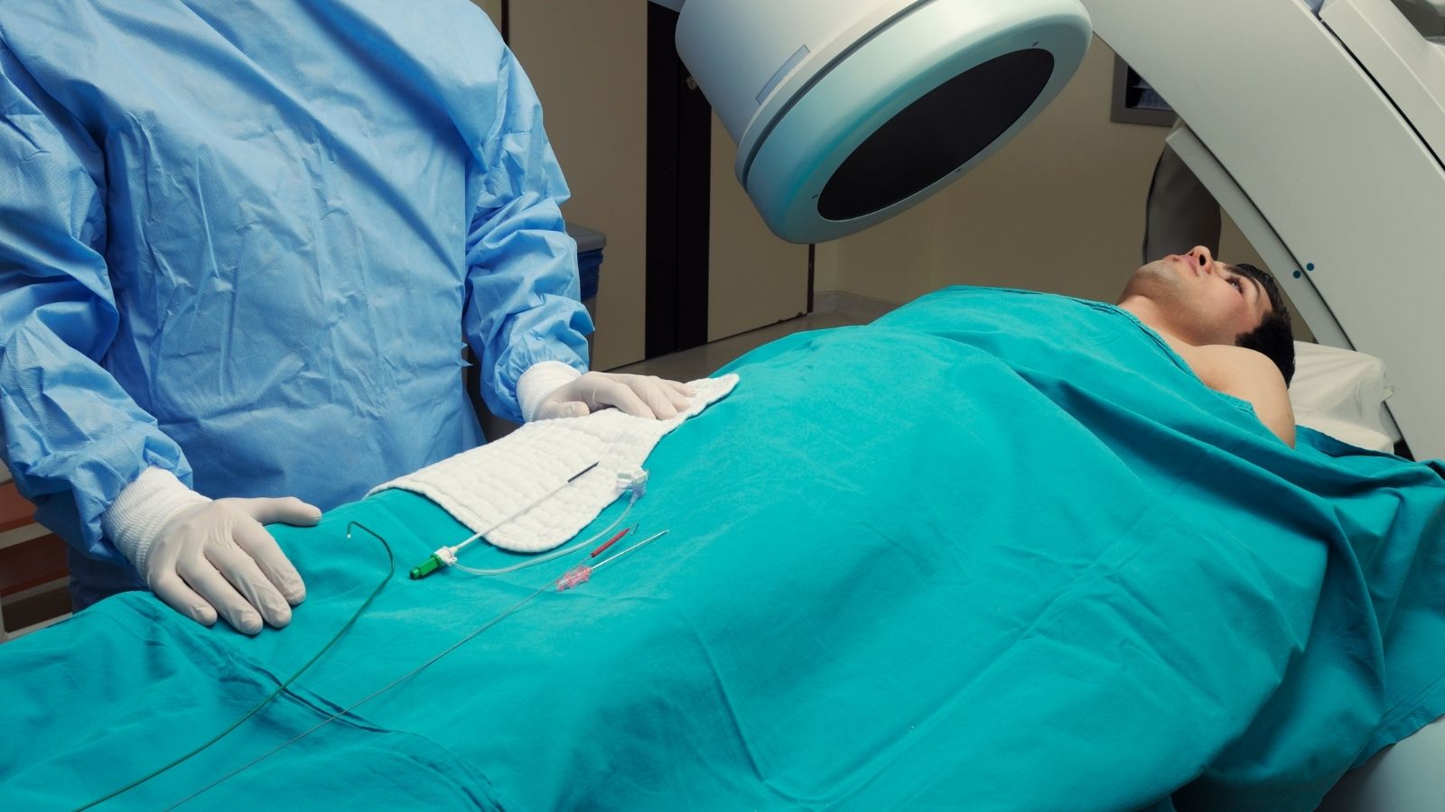
TAVI, which emerged through advances in medical technology, is an interventional method that allows the aortic valve to be replaced without opening the chest or stopping the heart. Rather than a surgical operation, it is a catheter-based procedure performed in angiography laboratories. Biological leaflets obtained from bovine or porcine pericardial tissue are integrated into flexible metal stents that can be folded inward. This new valve is compressed and placed inside a thin tube (catheter), then usually advanced through blood vessels in the groin to reach the heart. Once it reaches the location of the old valve, it is expanded and opened, pressing the calcified tissue toward the vessel wall and allowing it to assume its new function within seconds. Its minimally invasive nature substantially reduces the systemic stress caused by surgery.
What Preparations Are Made for the Aortic Valve Before the TAVI Procedure?
The safe performance of TAVI depends on millimeter-precise measurements and detailed planning carried out before the procedure. The most important tool during this preparation stage is multidetector computed tomography, which creates a three-dimensional map of the heart. Based on tomography data, the diameter and circumference of the aortic valve and the density of surrounding calcification are calculated. When deciding on the size of the new valve, the distance between the valve and the coronary arteries supplying the heart is also measured to assess the risk of these arteries becoming blocked when the valve expands. During the same scan, the groin and abdominal vessels through which the valve-carrying catheter will pass are examined. By checking the width of the vessels and their internal curves, a roadmap is established to perform the procedure through the safest route.
How Is TAVI Aortic Valve Replacement Performed Step by Step Without Opening the Chest?
TAVI is generally performed in specialized laboratories equipped with advanced imaging systems or in hybrid operating rooms. In many cases, the patient does not need to be completely anesthetized; local anesthesia combined with sedative medications may be sufficient to numb the treatment area. The main steps of the procedure begin with the insertion of a small needle into an artery in the groin. Soft guidewires advanced through the blood vessel toward the heart pass through the narrowed valve and reach the left ventricle. In some cases, a balloon is inflated beforehand at the valve location to stretch the narrowed area and create a passage.
The system carrying the folded prosthetic valve is then advanced over these wires until it reaches the level of the calcified valve. If a balloon-expandable valve is used, the heart rate is temporarily increased at the moment of full expansion to prevent the heart from ejecting blood forward and displacing the valve. During this brief period of rapid pacing, the balloon is inflated to position the valve. In self-expanding systems, the sheath is slowly withdrawn, allowing the valve to assume its shape through body temperature. Immediately after the valve is released, ultrasound and pressure measurements are used to check the function of the new valve. Once no problems are detected, the system is removed from the blood vessel, and the entry site in the groin is closed using special vascular closure devices without external sutures, bringing the procedure to completion.
How Does Recovery Progress After TAVI Aortic Valve Replacement?
Because the integrity of the breastbone is preserved and the patient is not connected to a heart-lung machine, recovery after TAVI proceeds quite rapidly. Patients are generally transferred to their beds while conscious following the procedure. After a few hours of rest, they are allowed to sit, eat, and gradually stand up and walk. Since the need to remain connected to a ventilator rarely arises, the likelihood of experiencing lung problems is lower. The absence of a large surgical wound allows patients to be discharged from the hospital walking independently after an average observation period of just one or two days. As there is no need to wait for bone healing, physical restrictions are kept to a minimum.
For Whom Is TAVI Aortic Valve Replacement a Suitable Option?
Initially considered a solution only for patients at high risk of undergoing surgery, TAVI now serves a much broader patient population thanks to technological advances. It has become a frequently preferred treatment method, particularly for individuals aged seventy and older whose anatomy is suitable for the procedure. According to current approaches, the patient groups receiving this treatment include:
- Elderly individuals
- Individuals with high surgical risk
- Patients in the intermediate-risk group
- Individuals whose chest anatomy is unsuitable for surgery
- Individuals with severe lung disease
- People with impaired kidney function
- Individuals who have previously undergone open-heart surgery
In these patient groups, the aim is to prevent the systemic exhaustion associated with open surgery, allowing the body to recover more quickly and enabling individuals to return to active life with much greater energy.
What Is the Valve-in-Valve Procedure and How Is It Performed on Patients Who Have Previously Undergone Aortic Valve Surgery?
In patients who previously received a biological valve through open-heart surgery, this valve may also become calcified and deteriorate over the years, just like natural valves. Performing a second operation using the same method on a patient whose chest has already been opened creates a difficult and risky process because of tissue adhesions. In such cases, the “Valve-in-Valve” method, which allows a new valve to be placed inside the old one without removing it, becomes an option. Using a catheter inserted through the groin, a new TAVI valve is positioned directly in the center of the deteriorated surgical valve. The new valve expands, attaches to the frame of the old valve, and immediately begins functioning. Thus, patients may have the opportunity to regain their health without experiencing the burden of a second open-heart surgery.
What Risks May Occur After TAVI and Aortic Valve Replacement?
Despite advances in medical technologies, it is known that these interventions performed through blood vessels also carry certain potential complications inherent to their nature. Possible situations that may arise during the procedure are carefully monitored. Some potential risks associated with interventional methods are listed below.
- Heart rhythm disorders
- Need for a temporary or permanent pacemaker
- Leakage around the valve edges
- Bleeding at the access site
- Blood vessel injuries
- Risk of blood clot embolization
Because the heart’s electrical conduction pathways are located very close to the aortic valve, pressure from the new valve on these pathways may cause the heart rate to slow down; this condition is generally managed by implanting a small pacemaker. Minor blood leakage around the valve edges can be corrected during the procedure using a balloon. To reduce the risk of blood clot embolization, additional safety measures may be implemented when necessary through filter systems that protect the cerebral blood vessels.
Which Medications Are Used After Aortic Valve Replacement?
After valve replacement, managing blood-thinning treatments is particularly important to prevent clot formation around the newly implanted valve. If a metallic (mechanical) valve has been implanted through open surgery, patients must take strong blood thinners regularly for life and undergo monitoring through blood tests. However, because valves implanted through TAVI are made from biological tissues, this requirement is more flexible. If the patient does not have another heart rhythm disorder, the long-term use of only one mild blood thinner (such as low-dose aspirin) may generally be considered sufficient. The type and dosage of medication to be used are planned in detail by the physician based on the patient’s clinical condition after the procedure.
How Long Does a Replaced Aortic Valve Last and When Can Patients Return to Daily Life?
One of the topics patients frequently wonder about is the durability of this valve implanted by advancing it through a blood vessel. Long-term follow-up of valves made from biological tissues has shown that they successfully maintain their structural integrity and can generally function without problems for more than ten years. This expected lifespan may vary from person to person due to the nature of biological valves. Returning to daily life is relatively straightforward because it does not require lengthy processes such as wound healing. During the first days after discharge, patients are advised to avoid strenuous physical activity and movements that may strain the groin area. Generally, after a short rest period of one or two weeks, patients can begin walking, travel, and comfortably return to their social lives.
What Should Be Considered to Protect Heart Health After Aortic Valve Replacement?
Although the mechanical burden on the heart has been reduced with a new aortic valve, maintaining overall heart health remains a lifelong responsibility. Individuals are advised to make certain adjustments to their daily habits so that the valve and the coronary arteries supplying the heart can maintain their healthy structure. The following measures should be considered to improve quality of life after the procedure:
- Limiting salt consumption
- Maintaining blood pressure control
- Keeping blood sugar levels balanced
- Completely avoiding tobacco products
- Taking regular walks at a gentle pace
- Adopting a Mediterranean-style diet
- Taking prescribed medications regularly
- Not neglecting outpatient follow-up appointments
These lifestyle changes substantially support the renewed valve in performing its function in harmony with the heart for many years. Taking care of physical health is the strongest factor in making the improvement achieved through medical intervention sustainable.

Prof. Dr. Kadriye Orta Kılıçkesmez is one of the leading figures in the field of Turkish cardiology. She was born on January 24, 1974, in Tekirdağ. After completing her undergraduate education at Istanbul University Cerrahpaşa Faculty of Medicine, she chose cardiology as her specialty and received her specialist training at the Cardiology Institute of the same university. In 2015, she was appointed by the university to establish the Şişli Etfal cardiology clinic and Angio laboratory. Becoming a professor in 2017, Kadriye Kılıçkesmez established the cardiology clinic and Angio laboratory of Prof. Dr. Cemil Taşçı Hospital in 2020 and ensured that the clinic became a training clinic.