Türkçe
 Français
 Deutsch
 Italiano
 Español
 العربية
 Русский

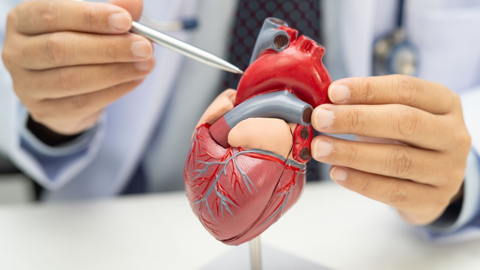
Mitral valve function and anatomy describe the tissue architecture and operating mechanism of the biomechanical valve system that maintains the flow of oxygenated blood inside the heart in one direction. This vital mechanism acts as a dynamic control point that prevents blood from leaking backward as it is pumped into the body.
Structurally, this region consists of flexible tissues that interlock with one another and specialized supporting connections that keep these tissues balanced with every heartbeat. This architecture, which flexes in synchronization with the rhythmic movements of the heart, instantly adapts to changing internal pressures.
At the functional level, the system’s primary task is to support the uninterrupted progression of circulation along the correct pathway. Thanks to the coordinated movement of the valve tissues, the amount of oxygen delivered to the organs is preserved, and the heart uses its energy efficiently.
When the integrity of this delicate system, which gradually wears down over time, deteriorates, circulatory balance can be restored through intravascular interventions using modern interventional cardiology approaches.
What Is the Mitral Valve and Where Is It Located in the Heart?
The heart is a powerful four-chambered pump that works day and night without stopping to deliver the oxygenated blood needed by the organs into the blood vessels. While the chambers on the right side send oxygen-depleted blood returning from the body to the lungs for oxygenation, the chambers on the left side distribute oxygenated blood arriving from the lungs to the organs. The mitral valve is located at the passage between the upper left chamber, called the left atrium, and the lower left chamber, called the left ventricle.
This structure can be imagined as a special door positioned between two rooms that opens in only one direction. It allows oxygenated blood in the upper chamber to flow easily into the lower chamber, while firmly closing its doors when the lower chamber contracts, largely preventing blood from flowing backward. Thus, the pumping force generated by the heart is not wasted, and blood is directed toward the tissues through the main artery, the aorta.
How Does Mitral Valve Function Contribute to Blood Circulation?
The left ventricle generates considerable pressure with each heartbeat to deliver blood from the brain to the tips of the toes. Mitral valve function comes into play precisely at this moment of contraction. As soon as the pressure inside the ventricle rises, the valve leaflets interlock, helping prevent blood from flowing back to where it came from, namely the left atrium and, from there, toward the lungs.
When the valve performs this function properly, the circulatory system operates in a balanced and efficient manner. However, if the valve cannot open sufficiently, blood cannot pass easily into the lower chamber; if it cannot close sufficiently, some of the pumped blood flows backward. In both situations, the heart must work much harder than normal to deliver the same amount of blood to the body. Therefore, the healthy functioning of this valve is one of the fundamental requirements for a person to walk without becoming tired and breathe comfortably in everyday life.
What Are the Main Structures That Make Up Mitral Valve Anatomy?
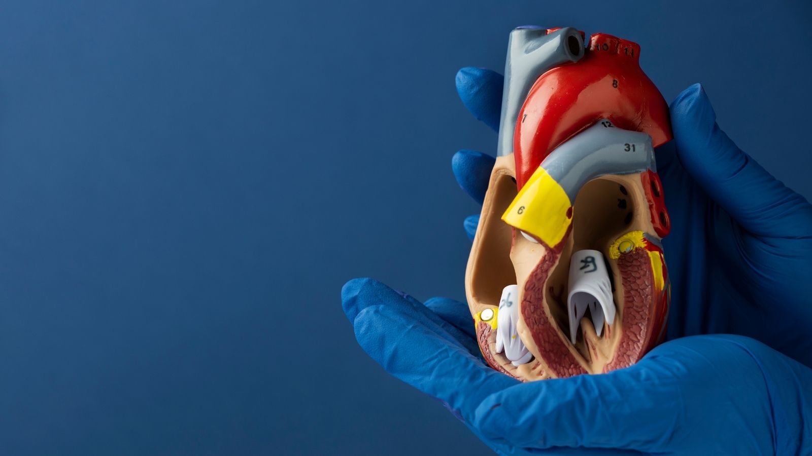
From a medical perspective, the mitral valve is not an independent tissue consisting of only two leaflets. In interventional cardiology and structural heart health, this region is evaluated as an integrated mechanism composed of directly interconnected parts. Just as coordination between a sailboat’s mast, sailcloth, and ropes is important, coordination between the supporting structures and components of the valve is equally critical.
The main components that make up mitral valve anatomy are:
- The flexible valve framework called the annulus
- The anterior and posterior valve leaflets
- Supporting fibers called chordae tendineae
- Papillary muscles connected to the heart muscle
- The walls of the left ventricle and left atrium
Calcification, loosening, elongation, or damage related to vascular obstruction that develops over time in any of these components can disrupt the valve’s closing balance and create conditions for various heart valve diseases.
What Is the Structure of the Annulus, the Framework of the Mitral Valve?
The annulus is a fibromuscular framework that anchors the base of the valve leaflets to the heart wall and provides the valve with a physical foundation. Although many people imagine this framework as a flat ring or circle, the annulus actually has a three-dimensional, curved structure. This special geometry, which resembles a saddle placed on a horse’s back, flexes with each heart contraction, reducing the mechanical load on the valve.
The anterior portion of the framework consists of very firm and durable connective tissue and is directly adjacent to the aortic valve, the heart’s main outlet. In contrast, the posterior portion is thinner, more flexible, and predominantly composed of muscular tissue. During cardiac enlargement or the progression of heart failure, the rigid anterior portion maintains its shape, while the flexible posterior portion may expand because it cannot withstand the pressure. Under normal circumstances, when the heart contracts, this framework reduces its area by approximately twenty to thirty percent, making it easier for the leaflets to meet. However, when the framework enlarges, this narrowing support decreases, and the leaflets may become unable to reach each other.
How Do Mitral Valve Leaflets Direct Blood Flow?
The two main tissue structures covering the valve opening are called the anterior and posterior leaflets. In a healthy adult, the passage area formed by these leaflets when open is approximately four to six square centimeters. The two leaflets have quite different structural characteristics, but when they close, they complement each other like pieces of a puzzle.
Although the anterior leaflet attaches to approximately one-third of the valve framework, it is relatively long and covers most of the total valve area. It also forms a boundary of the blood outflow passage from the left ventricle toward the aorta. The posterior leaflet, on the other hand, extends around a broad portion representing two-thirds of the framework but is shorter in length. The posterior leaflet has small natural indentations that divide it into three separate segments. Cardiologists evaluate these segments by numbering them to identify precisely which region of the valve has a problem. The corner points where the two leaflets meet at the sides are called commissures.
How Do the Mitral Valve Cords and Muscles Support the Valve?
Beneath the leaflets lies a very important support mechanism extending into the internal cavity of the left ventricle. The high pressure generated when the heart contracts attempts to push the leaflets toward the left atrium. The structures that prevent the leaflets from turning backward under this pressure are the chordae tendineae, which resemble parachute cords, and the papillary muscles to which these cords are anchored.
The supporting elements beneath the leaflets and their characteristics are:
- Primary chordae that hold the free edges
- Secondary chordae that reduce tension in the leaflet body
- Tertiary chordae that extend directly to the valve base
- The anterolateral papillary muscle supplied by two blood vessels
- The posteromedial papillary muscle supplied by one blood vessel
The details of this blood supply are clinically very important. Since the anterolateral papillary muscle receives blood from two different coronary arteries, it is relatively more resistant to vascular blockages. However, the posteromedial papillary muscle is usually supplied by a single branch originating from the right coronary artery. Therefore, during a heart attack affecting the inferior wall of the heart, this muscle can easily become deprived of oxygen and sustain damage.
How Does the Mitral Valve Function Biomechanically with Every Heartbeat?
Mitral valve biomechanics depend on coordination between the heart’s electrical stimulation and pressure changes between its chambers. When the heart muscle begins to relax, pressure inside the left ventricle rapidly decreases. As soon as ventricular pressure falls below left atrial pressure, the valve opens automatically. First, a rapid filling phase occurs, followed by a brief pause, and finally, the remaining blood is transferred into the lower chamber through atrial contraction. In a healthy valve, almost no significant resistance occurs during this passage, and the pressure difference remains below two millimeters of mercury.
When the ventricle fills and contraction begins, the internal pressure rapidly rises. While fluid movements and vortices created by the blood bring the leaflets toward each other, the papillary muscles also contract to keep the cords taut. For a healthy seal, it is not enough for the leaflets to merely touch at their tips; their edges must overlap along a surface measuring approximately five to eight millimeters. This secure contact area is called the coaptation reserve and represents the most important physical safeguard against backward blood leakage.
What Are the Main Diseases That Impair Mitral Valve Function?
Wear and tear developing over the years, previous infections, or other disorders affecting the heart muscle can disrupt this delicate biomechanical balance. When evaluating valve diseases, the first consideration is whether the problem originates from the valve tissue itself or from enlargement of the heart muscle. The mobility of the leaflets also plays a decisive role in treatment planning.
The main disease groups and movement abnormalities affecting mitral valve function are:
- Mitral stenosis, known as narrowing of the valve
- Primary mitral regurgitation caused by leaflet damage
- Secondary mitral regurgitation caused by cardiac enlargement
- Annular enlargement with normal leaflet movement
- Valve prolapse with excessive leaflet movement
Calcification and retraction that restrict leaflet movement
This classification helps guide decisions about whether the patient’s treatment should involve open surgery or an interventional procedure performed through the blood vessels.
What Causes Mitral Valve Stenosis and How Does It Strain the Heart?
Mitral valve stenosis is the narrowing of the opening through which blood passes as a result of the valve leaflets thickening, losing their flexibility, and adhering to each other at their junctions. The most common cause of this condition in the population is rheumatic heart disease resulting from throat infections experienced during childhood or adolescence that were not adequately treated. At older ages, extensive calcification developing in the valve framework can also cause stenosis.
When the valve area narrows, oxygenated blood arriving from the lungs cannot pass easily from the left atrium into the left ventricle. Difficulty in passage causes blood to accumulate in the upper chamber and increases the pressure there. Over time, the left atrium enlarges because it cannot withstand this pressure, and the heart’s normal electrical system may become disrupted, resulting in rhythm disturbances. The increased pressure in the upper chamber is transmitted backward, affecting the pulmonary vessels and creating conditions for fluid accumulation in the lungs.
What Causes Mitral Valve Regurgitation and Leakage?
Mitral valve regurgitation, commonly referred to as valve leakage, occurs when the leaflets cannot close completely during ventricular contraction, allowing blood to leak backward into the left atrium. This condition may arise from primary (structural) or secondary (functional) causes. In primary regurgitation, the problem lies directly in the valve tissue itself; the leaflets may stretch and prolapse toward the atrium because of connective tissue weakness, or one of the cords holding the valve may rupture. In secondary regurgitation, the leaflets themselves are actually intact, but enlargement of the heart muscle causes the papillary muscles to move apart, pulling the leaflets downward and preventing them from closing.
The main factors that may cause mitral valve leakage include:
- Mitral valve prolapse, meaning prolapse of the leaflets
- Barlow’s disease associated with connective tissue weakness
- Infective endocarditis affecting the heart valve
- Rupture or excessive elongation of the supporting chordal fibers
- Enlargement of the heart muscle developing after a heart attack
- Atrial enlargement due to long-standing rhythm disturbances
Regardless of the cause, the backward flow of some blood with each heartbeat increases the heart’s workload and eventually causes fatigue of the heart muscle.
What Symptoms Do Mitral Valve Diseases Cause in Daily Life?
Valve diseases often progress slowly, and the heart attempts to compensate for the condition over a long period. Therefore, patients may not experience noticeable complaints in the early stages, and the problem may only be detected through a murmur heard when the doctor listens to the heart during a routine examination. However, when stenosis or leakage exceeds a certain threshold, the body becomes unable to tolerate the increasing burden and begins to produce signals that restrict daily life.
Common symptoms of mitral valve diseases include:
- Shortness of breath when walking uphill or climbing stairs
- Rapid fatigue and weakness while performing daily activities
- Coughing and a sensation of air hunger when lying down at night
- Needing to sleep with an elevated pillow
- Irregular palpitations felt in the chest
- Swelling of the ankles and legs
The presence of these complaints indicates that the heart can no longer compensate for the existing valve problem on its own and that it is time for a medical evaluation.
What Risks Arise If Mitral Valve Problems Are Left Untreated?
A mechanical defect in the valve cannot return to its original anatomical condition on its own or merely through medication. Drug treatments help remove excess fluid from the body, regulate heart rate, and alleviate symptoms; however, they cannot widen a physically narrowed valve or repair a ruptured cord. When intervention is delayed, the pressure and volume burden on the left atrium and left ventricle may cause permanent changes in the structure of the heart.
The adverse conditions that may result from delayed treatment of mitral valve disorders include:
- Progressive left- and right-sided heart failure
- Persistent rhythm disturbance known as atrial fibrillation
- Formation of blood clots inside the heart chambers
- Risk of stroke caused by a clot traveling to the brain
- Pulmonary hypertension, known as high blood pressure in the lungs
- Permanent stiffening of the heart muscle and loss of contractile function
It is particularly important for patients to receive appropriate treatment before permanent stiffening of the heart muscle and high resistance in the pulmonary vessels become established.
Which Diagnostic Methods Are Used to Evaluate the Mitral Valve?
Detailed imaging methods are used to understand the severity of a valve disease and determine the most appropriate treatment strategy. In the first stage, a superficial cardiac ultrasound examination performed through the chest wall, known as transthoracic echocardiography, is used. This examination provides a general understanding of the valve’s condition and the heart’s contraction strength. However, if an interventional treatment through the blood vessels is planned, the valve must be visualized much more closely and in three dimensions.
The methods used to diagnose mitral valve diseases and prepare for procedures include:
- Transthoracic echocardiography performed through the chest
- Two-dimensional and three-dimensional echocardiography performed through the esophagus
- Electrocardiography to evaluate heart rhythm
- Multislice cardiac computed tomography
- Catheterization procedures to measure pressures inside the heart
In particular, transesophageal echocardiography (TEE) provides a very clear view of the mitral valve because the esophagus lies immediately behind the heart. This method allows detailed examination of which leaflet segment is leaking, the extent of calcification, and whether any blood clots are present inside the heart.
How Is Mitral Valve Structure Measured Before Interventional Procedures?
The success of catheter-based treatments in interventional cardiology depends directly on measurements made with millimeter-level precision before the procedure. For example, in a patient with rheumatic mitral stenosis, a special assessment called the Wilkins score is performed to determine whether balloon treatment is appropriate. In this evaluation, leaflet mobility, thickness, the condition of the subvalvular cords, and the amount of calcification are each graded from one to four points. While a total score of eight or less indicates anatomy highly favorable for balloon treatment, scores above twelve, the presence of a clot inside the heart, or accompanying severe valve regurgitation make the balloon procedure unsuitable.
Similarly, in patients for whom a clipping procedure is planned to treat valve leakage, the depth of leaflet attachment, the length of leaflet contact, and the existing valve opening area are carefully measured. A valve area greater than four square centimeters before the procedure is an important safety criterion for preventing unwanted valve narrowing after the clip is placed.
In Which Situations Can Open Surgery Be Risky in Mitral Valve Treatment?
Traditional open-heart operations are an important treatment option that has been successfully performed for many years in numerous young individuals and people without additional illnesses. However, open surgery requires cutting and opening the breastbone, temporarily stopping the heart, and connecting the patient to a heart-lung machine. This process represents significant physical stress and a considerable recovery burden for the body.
The life-threatening risks of open surgery increase considerably in elderly individuals, heart failure patients whose heart contraction strength has significantly weakened, those who have previously undergone open-heart surgery, and people with chronic lung, kidney, or liver diseases. In such situations, a committee called the Heart Team, consisting of cardiologists and cardiac surgeons, directs the patient toward catheter-based methods offered by interventional cardiology to protect them from the substantial burden of surgery.
What Types of Solutions Does Interventional Cardiology Offer for Mitral Valve Diseases?
Interventional cardiology is a modern medical field that makes it possible to intervene in heart valves without opening the chest or stopping the heart, using only vascular access through the groin. Much like performing angiography, thin catheter tubes are advanced through the blood vessels to reach the right atrium. From there, a safe passage is created through the thin partition between the atria (fossa ovalis), providing direct access to the mitral valve.
The main nonsurgical mitral valve treatments performed by interventional cardiology specialists include:
- Percutaneous mitral balloon valvuloplasty
- Transcatheter edge-to-edge valve repair and clipping
- Implantation of a new valve inside an old biological valve
- Implantation of a valve inside a surgical repair ring
- Implantation of a valve in a severely calcified native valve bed
All these methods are performed in real time on the beating heart under the guidance of advanced X-ray equipment and three-dimensional ultrasound imaging through the esophagus.
How Is Balloon Dilation Performed for Mitral Valve Stenosis?
Percutaneous mitral balloon valvuloplasty (PMBV) is the first-choice interventional method for patients with rheumatic mitral valve stenosis who have suitable anatomy. In this procedure, the femoral vein in the groin is accessed, and a special guidewire is used to cross into the left atrium. A specially designed Inoue balloon, which can be inflated gradually, is then positioned precisely in the center of the narrowed valve.
First, the distal portion of the balloon is inflated and positioned beneath the valve. Immediately afterward, its middle and upper portions are briefly inflated, mechanically separating the fused valve corners (commissures) toward either side. After this inflation, which lasts only a few seconds, the balloon is deflated and withdrawn. At the end of the procedure, the aim is to increase the narrowed valve area to more than one and a half square centimeters and rapidly reduce the elevated pressure inside the left atrium. In the vast majority of appropriately selected patients, this procedure significantly alleviates symptoms and can postpone the need for open surgery by approximately ten to twenty years.
How Is the Nonsurgical Clipping Method Performed for Mitral Valve Regurgitation?
Transcatheter edge-to-edge repair (TEER) is a prominent option for patients with mitral valve regurgitation who face a high risk from open surgery or continue experiencing symptoms despite drug treatment. This procedure, commonly known as the clipping method, uses advanced devices such as MitraClip, Pascal, or similar technologies. The basic principle of this method is to bring together and attach the central portions of the leaking anterior and posterior leaflets to control regurgitation.
The left atrium is reached using a steerable catheter system advanced through the femoral vein. Under three-dimensional ultrasound guidance, the clip is aligned with the area where leakage is greatest. The arms of the clip are opened and lowered into the ventricle, the leaflet edges are carefully grasped, and the clip is closed to secure the leaflets together. Thus, instead of one large opening, two smaller openings side by side, resembling the figure eight, are created through which blood can flow comfortably, while backward leakage is substantially reduced. Extensive scientific studies have demonstrated that this method significantly reduces hospitalizations and life-threatening risks in appropriately selected patients with valve regurgitation caused by heart failure.
How Is Mitral Valve Replacement Performed Through the Blood Vessels Without Open Surgery?
In some patients, the valve structure may not be suitable for clipping, extensive calcification may be present in the leaflets, or a biological valve surgically implanted years earlier may have worn out and deteriorated over time. In these individuals, for whom a second open-heart operation would be risky, transcatheter mitral valve replacement (TMVR), meaning valve replacement without open surgery, becomes an option. In this procedure, a new biological valve compressed inside a catheter is advanced through a blood vessel in the groin or through a small incision in the chest wall and placed inside the deteriorated old valve, surgical ring, or calcified native valve bed.
Important considerations before and during nonsurgical mitral valve replacement include:
- Measuring the internal diameter of the old biological valve
- Evaluating the amount of calcification for valve anchoring
- Performing a virtual valve simulation using computed tomography
- Examining the risk of narrowing of the left ventricular outflow tract
- Determining the appropriate access route through the groin or the apex of the heart
Virtual simulation performed using computed tomography before the procedure is particularly valuable. It allows advance calculation of whether the newly implanted valve will push the existing anterior leaflet and narrow the heart’s main outflow tract (LVOT), thereby improving procedural safety.
What Advantages Do Interventional Mitral Valve Treatments Offer Patients?
The most important benefit of interventional cardiology methods for patients is their minimally invasive nature, which preserves the body’s structural integrity. Avoiding cutting the breastbone and stopping the heart considerably facilitates recovery, particularly in elderly individuals or those with additional illnesses. After the procedure, the duration of intensive care stays is reduced, and patients can readjust to their daily lives much earlier.
The main advantages of catheter-based mitral valve interventions include:
- No large surgical incision in the chest
- No need for connection to a heart-lung machine
- Low risk of bleeding and wound infection
- Very low levels of pain after the procedure
- Hospital stays limited to a few days
- Rapid return to daily life and social activities
Thanks to these advantages, many patients who previously had no opportunity to undergo an intervention because of high risks can now enjoy a better quality of life by largely overcoming limiting symptoms such as shortness of breath and fatigue.
What Should Be Done to Protect Mitral Valve Health and Avoid Delays in Treatment?
Mitral valve anatomy and function interact directly with all other parts of the heart. If deterioration beginning in one component of the valve is not recognized and managed in time, it may initiate a chain of damage extending from the left atrium to the lungs and then to the right side of the heart. Therefore, it is necessary both to pay attention to general measures aimed at preserving valve health and to avoid missing follow-up appointments when a valve disease is already present.
The main considerations for supporting heart and mitral valve health include:
- Timely treatment of throat infections accompanied by fever
- Regular oral and dental health examinations
- Keeping blood pressure and cholesterol levels under control
- Limiting salt consumption according to the doctor’s recommendations
- Seeking medical examination without delay when shortness of breath or palpitations occur
- Regular echocardiographic monitoring of existing valve diseases
Appropriate evaluations performed before permanent damage develops in heart tissue directly affect treatment success. Today, thanks to advanced methods offered by interventional cardiology, such as balloon dilation, clipping, and intravascular valve replacement, valve function can be supported again in suitable patients without the need for open surgery, helping secure the future health of the heart.

Prof. Dr. Kadriye Orta Kılıçkesmez is one of the leading figures in the field of Turkish cardiology. She was born on January 24, 1974, in Tekirdağ. After completing her undergraduate education at Istanbul University Cerrahpaşa Faculty of Medicine, she chose cardiology as her specialty and received her specialist training at the Cardiology Institute of the same university. In 2015, she was appointed by the university to establish the Şişli Etfal cardiology clinic and Angio laboratory. Becoming a professor in 2017, Kadriye Kılıçkesmez established the cardiology clinic and Angio laboratory of Prof. Dr. Cemil Taşçı Hospital in 2020 and ensured that the clinic became a training clinic.