Türkçe
 Français
 Deutsch
 Italiano
 Español
 العربية
 Русский

Mitral valve diseases are circulatory disorders that develop when the valve between the left chambers of the heart narrows or fails to close completely, allowing blood to leak backward. They mainly occur as mitral valve stenosis, mitral valve regurgitation, and mitral valve prolapse.
Causes of mitral valve diseases include previous infections accompanied by fever, age-related calcification, connective tissue weakness, and enlargement of the heart. These factors impair valve movement and increase the workload on the heart.
Treatment methods for mitral valve diseases include medication and monitoring, minimally invasive interventional cardiology procedures performed through blood vessels, and open surgery. Balloon dilation, clip-based repair, and catheter-based valve replacement performed without surgical incisions are modern approaches.
Catheter-based repairs planned according to the patient’s anatomical characteristics help regulate blood flow without stopping the heart. Timely interventional procedures help prevent progressive heart damage and preserve quality of life.
What Is the Mitral Valve and How Do Mitral Valve Diseases Begin in the Heart?
To understand how the heart works, it can be compared to a continuously operating water pump with four chambers. Oxygen-rich blood coming from the lungs first enters the upper left chamber of the heart, called the left atrium. From there, it passes into the lower left chamber, the left ventricle, to be pumped forcefully throughout the body. The structure located between these two chambers, which allows blood to flow in only one direction, is called the mitral valve. This valve consists of two flexible leaflets that open and close, a fibrous ring surrounding the valve, and thin cords resembling parachute strings that connect the leaflets to muscles in the heart wall.
With every heartbeat, these components move in coordination with one another. When the heart relaxes, the valve opens to allow blood to fill the lower chamber. When the heart contracts, the valve closes to prevent blood from flowing backward. Over time, if calcification, thickening, stretching, or rupture occurs in any of these components, the valve can no longer perform its function as effectively as before. Inadequate opening of the valve causes stenosis, while incomplete closure leads to backward leakage, known as regurgitation, initiating the disease process.
The main anatomical structures that form the mitral valve and ensure coordinated function include:
- Anterior and posterior valve leaflets
- Flexible annular ring surrounding the valve
- Chordae tendineae resembling parachute strings
- Papillary support muscles in the heart wall
- Transition area between the left atrium and ventricle
What Causes Mitral Valve Diseases and What Are the Risk Factors?
Processes that damage the structure of heart valves may sometimes progress silently over many years, while in other cases they may appear following a suddenly developing heart condition. Throat infections that were common in previous decades and were not adequately treated can leave behind stiffening and adhesions in valve tissue years later. Today, however, as life expectancy increases, age-related tissue degeneration and calcification have become much more common causes.
In addition, the valve tissue itself may have excessive flexibility from birth or weakness of the connective tissue. Conditions such as a previous heart attack, blockage of the coronary arteries, or high blood pressure that has remained uncontrolled for many years can also change the geometry of the heart and disrupt the valve’s closing mechanism. Furthermore, bacteria entering the bloodstream after tooth extraction or various surgical procedures may settle on the valve and cause damage to its tissue.
The main risk factors associated with the development of mitral valve diseases include:
- Rheumatic fever during childhood
- Age-related tissue calcification
- Congenital connective tissue laxity
- Previous heart attack and vascular blockage
- Long-standing uncontrolled high blood pressure
- Rhythm disorders that enlarge the heart chambers
- Microbial infections affecting valve tissue
What Is Mitral Valve Stenosis and How Does It Make the Heart Work Harder?
Mitral valve stenosis is a condition in which the valve between the left atrium and left ventricle loses its flexibility and cannot open sufficiently. In a healthy adult, the opening area of this valve is approximately four to six square centimeters, allowing blood to flow freely into the lower chamber without encountering an obstruction. However, when the valve leaflets become thickened, fuse together at their edges, or the fibers beneath the valve shorten and stiffen, this opening gradually narrows. When the valve area falls below two square centimeters, particularly below one and a half square centimeters, blood flow becomes significantly restricted.
Oxygen-rich blood that cannot pass through the narrowed opening begins to accumulate in the chamber behind it, the left atrium. This accumulation increases the pressure inside the left atrium and causes the chamber to enlarge over time. After a while, the elevated pressure is transmitted farther backward to the pulmonary vessels from which the blood arrives. Increased pressure in the blood vessels of the lungs creates an additional burden that makes breathing more difficult. If this process continues for many years, the increased pressure may also strain the right side of the heart and negatively affect the overall balance of circulation.
Changes that may develop in the heart and circulatory system as mitral valve stenosis progresses include:
- Significant increase in pressure within the left atrium
- Enlargement of the upper left heart chamber
- Blood congestion in the pulmonary veins
- Increased pressure in the pulmonary blood vessels
- Excessive strain on the right heart chambers
- Reduced amount of blood pumped to the body
What Are the Main Factors That Cause Mitral Valve Stenosis?
A large proportion of mitral valve stenosis cases are caused by acute rheumatic fever experienced during childhood or adolescence. Following a streptococcal throat infection, the body’s immune system may sometimes react against its own heart tissue. This initiates a slow process of healing and stiffening that continues for years. After twenty or thirty years without any symptoms, the edges of the valve become fused together, and the valve develops a narrow, funnel-like structure.
In older individuals, a different condition called degenerative stenosis may develop even without a history of rheumatic disease. With aging, calcium deposits, or calcification, begin to accumulate in the ring-shaped structure supporting the mitral valve. Over time, this calcification extends toward the bases of the leaflets, restricting the opening and closing movements of the valve. Although less common, congenital structural valve stenosis, previous radiation therapy to the chest, or gradual deterioration of previously implanted biological heart valves may also contribute to the development of stenosis.
The main factors that may cause mitral valve stenosis include:
- Valve adhesions caused by rheumatic fever
- Age-related calcification of the valve annulus
- Thickening and shortening of the fibers supporting the valve
- Congenital structural valve abnormalities
- History of radiation therapy to the chest
- Deteriorated older biological prosthetic valves
What Is Mitral Valve Regurgitation and How Does Leakage Develop in the Heart?
Mitral valve regurgitation is commonly described as leakage in a heart valve. When the lower left chamber of the heart contracts to send oxygen-rich blood throughout the body through the aorta, the mitral valve is expected to close tightly and ensure that blood flows only forward. If the valve leaflets cannot meet edge to edge or close properly against one another, some of the pumped blood leaks backward into the left atrium.
This backward leakage means the heart must perform additional work with every beat. The heart must both deliver the blood needed by the body and compensate for the volume of blood that leaks backward. During the early stages, the heart balances this situation by contracting more forcefully and enlarging its chambers slightly. Therefore, individuals may experience no complaints for a long time. However, this additional workload over many years can eventually cause the heart muscle to become fatigued, the chambers to enlarge further, and the heart’s contraction strength to decline.
The mechanical effects experienced by the heart during mitral valve regurgitation include:
- Backward leakage of oxygen-rich blood into the left atrium
- The heart working with excess blood volume during each beat
- Gradual enlargement of the left ventricular cavity
- Fatigue and thinning of the heart muscle walls
- Fluid overload transmitted to the pulmonary vessels
- Reduced effective blood flow to the organs
What Are the Differences Between Primary and Secondary Mitral Valve Regurgitation?
To develop an appropriate treatment plan for mitral valve regurgitation, it is necessary to distinguish whether the leakage originates from the valve itself or from the overall structure of the heart. In primary, or organic, mitral valve regurgitation, the problem lies directly in the valve leaflets or the string-like supporting cords. Mitral valve prolapse, in which the leaflet tissue is loose and bulges toward the left atrium when the heart contracts, is one of the best-known examples. Sometimes one of the thin cords supporting the valve may suddenly rupture, or an infection may damage a valve leaflet.
In secondary, or functional, mitral valve regurgitation, the valve leaflets themselves are actually intact. The main problem is enlargement of the heart chambers surrounding the valve. When the left ventricle becomes enlarged and more spherical because of a previous heart attack or heart muscle disease, the muscles supporting the valve from below are pulled sideways and downward. Because of this pulling, known as tethering or tenting, the otherwise healthy leaflets cannot meet in the middle. Similarly, enlargement of only the left atrium and the valve annulus due to long-standing rhythm disorders can cause the leaflets to move apart and lead to leakage.
The main clinical conditions associated with different types of mitral valve regurgitation include:
- Mitral valve prolapse and leaflet displacement
- Rupture of the chordae tendineae supporting the valve
- Left ventricular enlargement after a heart attack
- Valve tethering associated with heart failure
- Enlargement of the valve annulus caused by rhythm disorders
- Valve laxity associated with connective tissue diseases
What Symptoms Do Mitral Valve Diseases Cause in Daily Life?
When mitral valve diseases are mild or moderate, the body can compensate for the condition, allowing individuals to continue their daily activities without limitations. Symptoms generally appear when stenosis or regurgitation exceeds a certain threshold and pressure in the lungs begins to increase. The earliest and most commonly noticed symptom is shortness of breath during activities that were previously performed comfortably, such as walking, climbing stairs, or walking uphill.
As the disease progresses, shortness of breath may occur during lighter movements or even at rest. Many people report difficulty breathing when lying flat at night, forcing them to sleep with two or three pillows under their heads. Inadequate delivery of oxygen-rich blood to the body causes muscles to tire quickly and produces a feeling of weakness throughout the day. In addition, enlargement of the left atrium may affect the heart’s electrical conduction and cause palpitations described as fluttering like a bird in the chest or skipped heartbeats.
Symptoms frequently observed in daily life in people with mitral valve diseases include:
- Shortness of breath occurring early during physical exertion
- Breathlessness that awakens the person from sleep at night
- Need to sleep with multiple pillows
- Rapid fatigue and exhaustion during daily activities
- Sensation of irregular heartbeats and palpitations
- Swelling in the ankles and legs
- Persistent dry cough that worsens when lying down
What Problems Can Mitral Valve Diseases Cause If Left Untreated?
Advanced mitral valve diseases that remain untreated despite the appearance of symptoms may eventually lead to changes in the heart and lungs that are difficult to reverse. As the left atrium enlarges under continuous pressure and volume overload, the heart’s normal rhythm may become disrupted, leading to an irregular rhythm disorder called atrial fibrillation. When this rhythm disorder develops, blood flow within the atrium slows, increasing the likelihood of blood clots forming in the chamber’s recessed areas.
If these clots break loose and enter the bloodstream, they may cause serious conditions such as blockage of blood vessels in the brain. On the other hand, if elevated pressure in the pulmonary vessels persists for a long time, the structure of these vessels becomes stiffened, and pulmonary hypertension may become permanent. After a while, this condition may also strain the right side of the heart, which pumps blood to the lungs, causing swelling in the legs, fluid accumulation in the abdomen, and worsening signs of general heart failure. For this reason, intervention at the appropriate time, before the heart muscle and pulmonary vessels become exhausted, is extremely important.
Possible conditions that may develop when treatment of mitral valve diseases is delayed include:
- Persistent irregular heart rhythm
- Formation of blood clots inside a heart chamber
- Risk of stroke caused by clots entering the circulation
- Permanent increase in pulmonary vascular pressure
- Weakening of the heart muscle’s contraction strength
- Right-sided heart failure and swelling in the body
How Are Mitral Valve Diseases Diagnosed and Which Tests Are Performed?
The evaluation of mitral valve diseases begins with a detailed medical history and a physical examination using a stethoscope. When listening to the heart, sounds called murmurs, produced by blood passing through a narrowed valve or leaking backward, provide the first indication of a valve problem. The primary method used for definitive evaluation and grading is transthoracic echocardiography, or heart ultrasound, performed through the chest wall. This painless examination allows detailed measurement of leaflet movement, valve opening area, the volume of blood leaking backward, and the dimensions of the heart chambers.
When catheter-based interventional treatment is planned, the valve anatomy must be examined much more closely. At this stage, transesophageal echocardiography is performed using a thin probe advanced through the esophagus. Because the esophagus is located immediately behind the heart, this method provides clear three-dimensional images of the mitral valve. Furthermore, if the valve has extensive calcification or catheter-based implantation of a new valve is being considered, multislice computed tomography is performed to calculate the distribution of calcium and the heart’s internal measurements with millimeter-level precision.
The main methods used in the diagnosis of mitral valve diseases and preparation for procedures include:
- Detailed cardiac examination using a stethoscope
- Electrocardiography to assess heart rhythm
- Standard echocardiography performed through the chest wall
- Three-dimensional echocardiography performed through the esophagus
- Multislice contrast-enhanced cardiac computed tomography
- Tests measuring changes in valve pressure during exertion
Why Is Interventional Cardiology Preferred in the Treatment of Mitral Valve Diseases?
For many years, open-heart surgery was the standard treatment for patients with severe mitral valve stenosis or regurgitation. During open surgery, the heart is accessed by cutting the breastbone or entering between the ribs, the patient is connected to a heart-lung machine, and the heart is stopped so the valve can be repaired or replaced. Although surgical methods provide highly successful results in younger individuals without additional diseases, surgical risk increases considerably in older patients, individuals with reduced cardiac contraction strength, those with kidney or lung diseases, or patients who have previously undergone open-heart surgery.
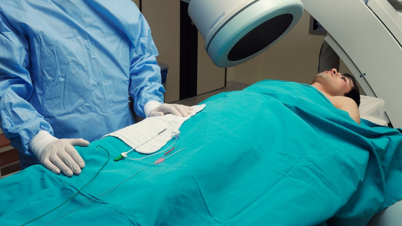
Catheter-based treatments developed in interventional cardiology offer a particularly valuable alternative for patients in these circumstances. These procedures are performed through veins in the groin or neck without opening the chest, stopping the heart, or requiring a heart-lung machine, thereby avoiding major surgical trauma to the body. In patients assessed by the Heart Team, consisting of cardiology and cardiovascular surgery specialists, and considered to have a high surgical risk, interventional methods help control symptoms and allow a return to daily life within a very short time.
The main advantages of interventional mitral valve treatments compared with surgery include:
- No incision in the breastbone
- Performed on the beating heart without stopping it
- No need for a heart-lung machine
- Can be performed safely in selected older patients and those with additional diseases
- Shorter intensive care and hospital stays
- Return to daily physical activities within a few days
- Lower risk of major surgical bleeding and infection
How Is Balloon Dilation Performed for Mitral Valve Stenosis?
In patients with severe rheumatic mitral valve stenosis who experience shortness of breath, percutaneous mitral balloon valvuloplasty is the preferred first-line interventional method when the valve structure is suitable. Before this procedure can be performed, echocardiography is used to assess the valve’s flexibility, thickness, and degree of calcification, and a suitability score is calculated. If the valve does not have excessive calcification, there is no clot inside the left atrium, and the stenosis is not accompanied by severe valve regurgitation, the patient is considered a highly suitable candidate for balloon treatment.
The procedure is generally performed without putting the patient fully to sleep, using local anesthesia in the groin area and mild sedative medications. A thin catheter is introduced through a vein in the groin and advanced to the right atrium of the heart. A special needle is then used to cross the thin septum between the right and left atria, providing access to the left atrium. A specially designed, hourglass-shaped Inoue balloon is positioned across the narrowed valve and gradually inflated. The inflated balloon separates the fused valve edges and enlarges the narrowed opening. Following the procedure, the valve area nearly doubles in the great majority of patients, providing rapid relief from shortness of breath, and this improvement may continue for many years.
The main characteristics of balloon dilation for mitral valve stenosis include:
- Access through the groin vein without a surgical incision
- Generally no need for general anesthesia
- Separation of fused valve edges using a balloon
- Rapid enlargement of the narrowed valve area
- Immediate improvement in breathing after the procedure
- Positive clinical effects that may continue for many years
How Is Non-Surgical Clip Repair Performed for Mitral Valve Regurgitation?
The most widely used catheter-based treatment developed for patients with mitral valve regurgitation who have a high risk from open surgery is transcatheter edge-to-edge repair, commonly known as the clip or clipping method. This technique is the catheter-based version of the surgical principle of suturing the leaflet edges together. It is performed by entering through a vein in the groin and advancing into the left atrium. For patient comfort and clear three-dimensional ultrasound imaging, the procedure is carried out under general anesthesia with guidance from transesophageal ultrasound and X-ray imaging.
A small clip system made from a special alloy, located at the tip of the catheter, is directed toward the edges of the anterior and posterior mitral valve leaflets that fail to close completely and allow blood to leak. At the point where leakage is greatest, the two leaflets are captured between the arms of the clip and secured together. In this way, the single wide opening that allows leakage through the center is transformed into a double-orifice structure through which blood can pass freely at the sides while backward leakage is largely prevented. When necessary, more than one clip may be implanted during the same session. Clinical studies have demonstrated that this method reduces hospital admissions and improves quality of life in both primary regurgitation caused by valve prolapse and secondary regurgitation associated with heart failure when symptoms persist despite medical treatment.
The main benefits of clip-based treatment for mitral valve regurgitation include:
- Mechanical joining of the leaking leaflet edges
- Significant reduction in the volume of backward blood leakage
- Precise application under three-dimensional ultrasound guidance
- Reduced hospital admissions due to heart failure
- Relief from shortness of breath and rapid fatigue
- Safe repair in patients who are not suitable for open surgery
What Are Catheter-Based Mitral Valve Replacement and Annular Reduction Methods?
In some patients, the mitral valve is so heavily calcified or the leaflet structure is so severely damaged that neither balloon dilation nor clip repair is sufficient. Similarly, a biological valve implanted during previous open surgery or a surgical repair ring may lose its function over the years. In such situations, where a second open-heart operation carries a high risk, catheter-based mitral valve replacement, known as transcatheter mitral valve replacement, may be performed. A new biological valve, advanced in a folded state through a vessel in the groin, is expanded and positioned directly inside the calcified native valve or deteriorated surgical valve.
Before this procedure, three-dimensional simulations are performed using computed tomography to calculate in detail whether the new valve might narrow the heart’s blood outflow tract. If a risk of outflow tract narrowing is identified, protective catheter techniques may be performed before or during the procedure to relieve the outflow tract and improve safety. In another interventional method called indirect annuloplasty, a special nitinol device is inserted through a neck vein into the coronary sinus, a vessel passing immediately outside the mitral valve. This device tightens the valve annulus from the outside, reducing the size of the enlarged valve and helping the leaflets move closer together again.
Other catheter-based methods used in advanced interventional mitral valve treatment include:
- Implantation of a new valve inside a deteriorated biological valve
- Catheter-based valve implantation inside a surgical repair ring
- Transcatheter valve implantation in a severely calcified native valve
- Reduction of the valve annulus through tightening from within the coronary sinus
- Preparatory protective procedures to preserve the heart’s outflow tract
What Is the Recovery Process Like After Interventional Mitral Valve Treatment?
Recovery after catheter-based mitral valve procedures is generally much faster and more comfortable than recovery following traditional open-heart surgery. Because the breastbone is not cut, patients do not face difficult recovery periods involving weeks of bone-healing pain or respiratory ventilator support. After the procedure, patients are usually transferred to a regular hospital ward following a short observation period, and the great majority can be discharged walking within twenty-four to forty-eight hours. The small vascular access site in the groin or neck closes naturally within a few days.
After discharge, patients can comfortably return to daily activities at home within a few days. Unlike open-heart operations involving mechanical valve implantation, catheter-based valve repairs generally do not require lifelong use of strong anticoagulants that necessitate strict blood-test monitoring. Instead, blood-thinning medications and cardiac drugs are used for a specified period according to the physician’s plan. To maintain recovery and allow the heart to adapt to its new condition, the most important steps supporting long-term treatment success include attending regular echocardiography examinations, monitoring blood pressure and heart rhythm, balancing salt intake, and maintaining good oral and dental health.
Recommended practices for a healthy recovery after interventional mitral valve treatment include:
- Avoiding strain on the groin access site during the first few days
- Taking prescribed cardiac and blood-thinning medications regularly
- Attending echocardiography follow-up appointments on the scheduled dates
- Avoiding excessive salt consumption in the daily diet
- Using preventive antibiotics before dental treatments
- Developing a habit of regular walking without overexerting the body

Prof. Dr. Kadriye Orta Kılıçkesmez is one of the leading figures in the field of Turkish cardiology. She was born on January 24, 1974, in Tekirdağ. After completing her undergraduate education at Istanbul University Cerrahpaşa Faculty of Medicine, she chose cardiology as her specialty and received her specialist training at the Cardiology Institute of the same university. In 2015, she was appointed by the university to establish the Şişli Etfal cardiology clinic and Angio laboratory. Becoming a professor in 2017, Kadriye Kılıçkesmez established the cardiology clinic and Angio laboratory of Prof. Dr. Cemil Taşçı Hospital in 2020 and ensured that the clinic became a training clinic.