Türkçe
 Français
 Deutsch
 Italiano
 Español
 العربية
 Русский

The most fundamental finding that distinguishes a heart attack from a panic attack and palpitations is the nature and duration of the pain. During a heart attack, a person experiences intense pressure in the center of the chest, resembling being squeezed in a vise, radiating to the left arm or jaw, not relieved by rest, and lasting longer than twenty minutes. This condition is usually accompanied by ice-cold sweating and nausea.
During a panic attack, a sharp, stabbing, needle-like pain is felt in the chest. This condition, whose intensity changes with breathing, is often accompanied by numbness in the hands, and the symptoms usually ease spontaneously within twenty minutes. In heart palpitations caused by an arrhythmia, rather than pain, the predominant sensation is a sudden, powerful fluttering in the chest, resembling the flapping of a bird’s wings, or the feeling of a skipped heartbeat.
Because these symptoms can imitate one another, making a definitive diagnosis at home is impossible. To rule out life-threatening risks, an ECG and high-sensitivity blood tests must be performed in the emergency department. In suspicious cases, the coronary arteries must be examined promptly through coronary angiography.
Why Are the Symptoms of Chest Pain and Heart Palpitations So Similar?
The way the human body perceives pain and discomfort signals originating from internal organs is much more different and complex than the way we perceive a needle piercing our skin. The heart muscle, lungs, esophagus, and muscles of the chest wall all transmit signals to the brain through the same nerve pathways in the spinal cord. While processing these signals from the internal organs (visceral organs), our brain may occasionally experience a kind of confusion regarding their origin. The pain caused by oxygen deprivation in the heart and the tension produced in the chest muscles by excessive adrenaline released by the brain during intense anxiety converge at the same nerve junctions in the spinal cord. When interpreting this complex flow of signals, the brain cannot identify the precise location of the pain. Consequently, it perceives the pain not only over the heart but as a broad area of discomfort felt in the left arm, shoulders, neck, back, or lower jaw. Furthermore, our nervous system pumps adrenaline into the bloodstream whenever it perceives danger, whether that danger is a blocked artery or simply the triggering of a phobia. This causes completely different illnesses and conditions to activate the same alarm signals in the body.
What Happens Physically to Our Bodies During a Panic Attack?
A panic attack is an extremely powerful but false alarm sent by the mind to the body when there is no actual physical threat or organic disease. During this process, the amygdala, located deep within the brain and responsible for regulating fear, is suddenly activated. This activation causes enormous amounts of adrenaline and noradrenaline to be released from the adrenal glands into the bloodstream. These hormones circulating through the blood vessels stimulate the heart’s natural pacemaker, causing the heart to beat much faster and blood pressure to rise suddenly. During this time, the person instinctively begins breathing rapidly and deeply. This rapid breathing, known medically as hyperventilation, causes the amount of carbon dioxide in the blood to decrease rapidly. The reduction in carbon dioxide disrupts the chemical balance of the blood and temporarily lowers calcium levels. This is the main reason for the unusual numbness and tingling felt in the hands, feet, and around the lips. Severe contractions of the chest muscles cause sharp, stabbing pains that feel like a knife piercing the chest and vary in intensity with breathing.
The physical reactions most frequently described by patients during a panic attack include:
- Palpitations
- Sweating
- Trembling
- Numbness
- Tingling
- Shortness of breath
- Stabbing chest pain
These symptoms usually intensify like an avalanche within a few minutes, reaching their peak. Within approximately half an hour, as the body’s adrenaline reserves are depleted, they gradually subside and are replaced by profound fatigue. The pain usually remains in the chest area and does not tend to spread widely as it does in an actual arterial blockage.
What Actually Happens in Our Coronary Arteries During a Heart Attack?
The condition commonly known as a heart attack is not a medical false alarm or a psychological manifestation, but the result of a completely real, physical, and mechanical blockage. Over years of unhealthy eating, smoking, diabetes, high blood pressure, and genetic influences, cholesterol deposits (plaques) form on the inner surfaces of the coronary arteries that supply the heart. Over time, these plaques may become inflamed like a pimple and suddenly rupture. Our body perceives this rupture as a wound and gathers platelets in the area to stop the bleeding. However, this defensive mechanism creates a blood clot that completely blocks the already narrowed artery. Within seconds, that region of the heart muscle is deprived of blood and oxygen. The oxygen-deprived tissue begins producing waste substances such as lactic acid, which strongly stimulate the nerve endings surrounding the blood vessels. The resulting pain is not a needle-like, stabbing pain or one that changes with breathing, as in a panic attack. Instead, it creates a crushing, squeezing sensation, as though an enormous weight has settled directly in the center of the chest, a stone is resting on it, or the rib cage is being compressed in a vise.
Some characteristic physical symptoms of a heart attack include:
- Chest pressure
- Cold sweating
- Nausea
- Vomiting
- Jaw pain
- Left arm pain
- Back pain
These complaints do not improve with rest or relaxation; they become more severe over time and last longer than twenty minutes. The accompanying ice-cold, clammy sweating and intense fear of death are among the clearest indicators that the body is experiencing serious physical damage.
How Do Arrhythmias, Commonly Known as Heart Palpitations, Develop?
Pathological heart palpitations, which differ greatly from the stress-related acceleration seen during panic attacks, originate from malfunctions in the heart’s own electrical system. Our heart has an extraordinary generator and wiring system that initiates every contraction. This natural generator, located in the right atrium, determines the heart’s rhythm by producing regular electrical signals at an average rate of sixty to one hundred per minute. However, during rhythm disorders (arrhythmias), certain abnormal groups of cells outside this main generator suddenly take control and begin producing electrical signals independently at extremely high speeds. Sometimes additional electrical pathways (accessory pathways) that have existed in the heart since birth but become active later create short circuits. These short circuits can suddenly increase the heart rate within seconds to one hundred and fifty or even two hundred and fifty beats per minute. The heart begins beating so rapidly that it contracts again before it even has time to fill with blood. A heart contracting while insufficiently filled cannot pump enough blood to the body or its own vessels. Therefore, even without any blockage in the major arteries, blood pressure falls and the heart muscle becomes deprived of oxygen.
Sensations frequently experienced by patients during rhythm disorders include:
- Fluttering like a bird’s wings
- Skipped heartbeats
- Dizziness
- Darkening of vision
- Feeling faint
Unlike panic attacks, these episodes usually do not begin gradually; instead, they start suddenly, as though an electrical switch has been pressed, and end just as abruptly seconds or hours later.
Which Warning Signs Indicate That We Need to Go to the Emergency Department?
Because the symptoms of these three conditions frequently overlap, it is essential to recognize certain warning signs to distinguish harmless situations from life-threatening ones. A previous diagnosis of an anxiety disorder or panic attacks never guarantees that the chest pain a person is experiencing at that moment is unrelated to the heart. Individuals with psychological problems can develop heart diseases just like anyone else. If the sensation in the chest is not a stabbing or needle-like pain but crushing pressure spreading over a broad area, it should be taken seriously. Pain that increases with exertion, such as climbing stairs or walking, does not disappear with rest, and lasts longer than twenty minutes is one of the strongest danger signals.
Some high-risk patient groups who may present with atypical symptoms that make diagnosis more difficult include:
- Women
- Older adults
- People with diabetes
- Patients with kidney disease
Long-standing diabetes, in particular, can damage nerve endings, meaning these individuals may never experience the classic crushing chest pain. Instead, they may only experience sudden, profound shortness of breath, persistent burning in the stomach, extreme unexplained fatigue, or darkening of vision accompanied by cold sweating. Temporary loss of consciousness (syncope) during or immediately after palpitations is also one of the most serious warning signs that the situation has progressed beyond simple excitement or a panic attack and that a dangerous process has begun in which the heart cannot pump blood adequately to the body.
Why Can ECGs and Similar Standard Tests Sometimes Be Insufficient for Diagnosis?
When a person visits an emergency department or outpatient clinic with chest pain or palpitations, the medical process usually begins with standard external (non-invasive) tests. The first step is always an electrocardiogram, or ECG. An ECG records the heart’s electrical activity at that particular moment on paper. During a panic attack, an ECG generally shows only an increased heart rate while the rhythm remains normal. During a heart attack, however, characteristic wave abnormalities indicating damage to the heart muscle may be detected. In addition, blood tests (cardiac troponin) are performed to detect certain proteins released into the bloodstream when heart muscle cells die.
Some basic examination methods frequently used in hospitals include:
- Electrocardiography
- Echocardiography
- Exercise stress test
- Holter monitor
- Blood tests
However, these tests have important limitations and restrictions. An ECG reflects only the few seconds during which it is recorded. If a person’s palpitations have stopped by the time they reach the hospital, the ECG may appear completely normal. Even Holter monitors worn for several days may sometimes fail to capture a brief episode of palpitations occurring only once a week. Although echocardiography, which uses ultrasound to examine the heart, shows the heart’s contraction strength and valves very clearly, it cannot visualize the interior of the coronary arteries with millimeter-level precision. An exercise stress test produces a positive result only when an arterial narrowing exceeds a certain critical threshold; it may overlook plaques at earlier stages or those creating hidden risks. It is precisely in these situations, when external examinations do not provide a clear answer, uncertainty remains, or immediate intervention is required, that the source of the problem must be investigated directly.
How Do Interventional Cardiology and Angiography Through the Wrist Work?
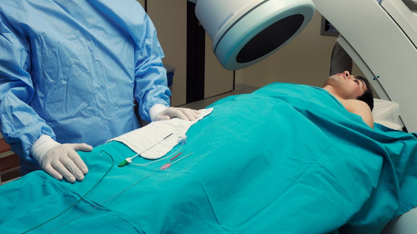
When external tests are insufficient, interventional cardiology is among the most powerful tools available to medicine for the definitive diagnosis and treatment of life-threatening blockages or rhythm disorders. Interventional cardiology is a specialized medical discipline that can reach deep inside the heart using only very thin vascular pathways, without opening the chest or making large surgical incisions. Coronary angiography is the gold standard for clearly visualizing the condition of the coronary arteries that supply the heart. In the past, this procedure was primarily performed through the large artery in the groin. Today, however, thanks to the possibilities offered by modern medicine and physicians’ experience, angiography procedures are largely performed using the transradial method, through the small artery in the wrist.
Some important advantages of angiography through the wrist for patients include:
- Early discharge
- Reduced bleeding
- Greater comfort
- Rapid mobilization
During the procedure, the patient remains fully awake, and only the wrist area is numbed using a small needle. Thin plastic tubes (catheters), approximately the thickness of spaghetti, are inserted through the wrist and follow the blood vessels until they reach the heart. A special contrast dye is then injected, and X-ray images are taken to map the arteries. While procedures performed through the groin require patients to lie on their backs for hours without moving their legs to prevent bleeding, the wrist approach allows patients to stand up immediately afterward, eat their meals, and, in many cases, walk home on the same day.
How Is a Stent Procedure Performed When an Arterial Blockage Is Detected?
If coronary angiography identifies a narrowing or complete blockage that seriously obstructs blood flow inside the coronary arteries and compromises the nourishment of this vital organ, the procedure can move from diagnosis to treatment within seconds. Particularly if the patient is experiencing a heart attack at that moment, rapidly opening the blocked artery becomes a race against time to save the heart. Extremely thin, specialized guidewires are advanced through the vessel to the blocked area. A miniature balloon in its deflated state is then guided over this wire and positioned directly in the center of the narrowing. When the balloon is inflated under high pressure, the calcified plaque blocking the vessel is compressed against the arterial wall, opening the passage. However, using a balloon alone may cause the artery to contract and close again shortly afterward. To prevent this, a special supporting structure called a stent is placed in the opened area.
A stent is a metallic, cage-like structure resembling the springs inside ballpoint pens, although it is much thinner. Positioned inside the artery on an inflated balloon, the stent remains as a permanent scaffold against the vessel wall after the balloon is deflated and removed, helping the artery stay open. Today, instead of bare-metal stents, advanced stents coated with special medications are used. These drug-eluting stents help significantly reduce the possibility of the inside of the stent narrowing again (restenosis) months or years later by suppressing excessive cell proliferation that may occur while the vessel heals. In suitable cases, special drug-coated balloons that release medication without leaving an additional metal structure inside the vessel are also successfully used during treatment.
How Can Unnecessary Procedures Be Avoided in Borderline Arterial Narrowings?
Coronary angiography does not always reveal clear situations in which an artery is completely blocked or severely narrowed. Sometimes the narrowing seen on the screen may appear to be between forty and seventy percent when assessed visually, placing it in a “borderline” category. Deciding to insert a stent based solely on visual judgment in such gray areas may expose the patient to an unnecessary procedure and medications they may need to use for life. Modern interventional cardiology uses advanced technologies to eliminate this uncertainty. A specialized technology called Fractional Flow Reserve (FFR) is used to determine whether a narrowing within an artery genuinely interferes with the heart’s blood supply.
Some advanced technological methods used during this process include:
- Fractional Flow Reserve (FFR)
- Intravascular Ultrasound (IVUS)
- Optical Coherence Tomography (OCT)
In the FFR method, a wire with a pressure sensor as thin as a human hair at its tip is passed beyond the narrowing inside the artery. Special medications are administered to stimulate the heart, and the pressure difference between the areas before and after the narrowing is measured. If the narrowing genuinely impairs blood flow at the cellular level, a decision is made to place a stent. Otherwise, monitoring the patient with medication and lifestyle changes is considered more appropriate. In addition, imaging systems that display the inside of the artery like a microscope (IVUS or OCT) can be used to examine the structure of the arterial wall, the cause of the narrowing, and whether the implanted stent fits properly against the vessel wall with millimeter-level precision. In this way, the reliability and durability of the treatment are improved.
What Role Does Catheter Ablation Play in Treating Heart Rhythm Disorders?
But what happens if the problem is not blocked arteries but faulty signals in the heart’s electrical system? Severe palpitations (arrhythmias) caused by additional electrical pathways or abnormal cellular foci inside the heart require lasting physical treatments, unlike psychologically triggered panic attacks. Electrophysiological studies (EPS) and catheter ablation are used to prevent these patients from needing lifelong medications for palpitations, end repeated emergency department visits, and eliminate the risk of sudden fainting. Like angiography, this procedure is performed in a catheterization laboratory, usually by accessing a vein in the groin.
Special wires fitted with small gold or platinum electrodes are placed inside the heart, allowing highly detailed ECG recordings to be obtained directly from within the heart with measurements accurate to milliseconds. Using computer systems, doctors send controlled electrical impulses to the heart to trigger, in the laboratory, the frightening palpitations that the patient experiences in everyday life. As soon as the palpitations begin, the faulty electrical circuit or additional pathway inside the heart is mapped and identified. Once the problematic area has been located with millimeter-level accuracy, ablation, meaning destruction of the tissue, is performed. Radiofrequency energy delivered through the tip of the ablation catheter heats a tiny point of abnormal tissue, making it incapable of conducting electrical impulses. If the focus causing the palpitations lies very close to the heart’s main electrical conduction network, cryoablation, a method that destroys tissue by freezing it, is preferred to prevent heat from damaging surrounding tissues. When performed in suitable patients, these procedures have the potential to achieve high success rates and provide long-term, lasting solutions for heart rhythm disorders.

Prof. Dr. Kadriye Orta Kılıçkesmez is one of the leading figures in the field of Turkish cardiology. She was born on January 24, 1974, in Tekirdağ. After completing her undergraduate education at Istanbul University Cerrahpaşa Faculty of Medicine, she chose cardiology as her specialty and received her specialist training at the Cardiology Institute of the same university. In 2015, she was appointed by the university to establish the Şişli Etfal cardiology clinic and Angio laboratory. Becoming a professor in 2017, Kadriye Kılıçkesmez established the cardiology clinic and Angio laboratory of Prof. Dr. Cemil Taşçı Hospital in 2020 and ensured that the clinic became a training clinic.